Category Archives: General paediatrics

Infantile spasms

Range of different shudders and twitches seen in babies and infants. Shudders and stereotypy can be dramatic, self stimulating behaviours, many kinds! Esp at 6-9/12, triggered by excitement but can also be boredom! So context important.  Can have a few together, but cluster would suggest epileptic.  Looks well. 

Differential is myoclonus, spasms (where at start may appear neurologically normal).

Infantile spasms are easy when classic, but are often atypical so can be difficult! 90% present before age 1, typically 3-7 months. Usually followed by upset so often confused with colic. Mostly when awake, arousal. Clusters of synchronous flexor spasms, usually of legs but possibly of head on torso. Definition is 0.5-2secs, so longer than jerk, some people think of an initial fast contraction followed by slower phase. Salaam attack is flexion of head and legs plus adduction of arms into midline. Rarely extensor, or mixed flexion of trunk and extension of limbs. Subtle presentations may just be chin. Can be unilateral.

Needs EEG to be sure, and even sleep EEG at that.  Low threshold since urgent treatment required.

Making diagnosis is urgent, as better initial control (in those without underlying aetiology) seem to have better developmental outcome). 

West syndrome – described by neurologist in his own son. Combination of spasms, developmental regression and hypsarrhythmia (see below). May initially be developmentally normal, become less visually attentive.  Flexion of head and trunk, arms extend/flex, abduct/adduct (can be asymmetrical if underlying hemiplegia).  Briefly upset or dazed, may be grimace.  On waking or falling asleep. Various Youtube videos available.

Rarely metabolic (consider PKU, Menkes, molybendum cofactors along with others). More often structural esp tuberosclerosis (TS – about 70% ) but also lissencephaly. Can be acquired eg post meningitis or congenital infection. Associated with Downs syndrome (actually more responsive to treatment).  Do Woods lamp and genetics. 20% cryptogenic.

Investigations

EEG by definition is abnormal, but in early phase standard awake EEG may be normal so consider sleep EEG.  Classical hypsarrhythmia in 50-70% = chaotic, high voltage.  But can be asymmetric.  If structural lesion, may be burst suppression (sudden high voltage then brief flattening) or focal features.

Treatment

ICISS (2016) showed combination of steroids and vigabatrin superior to either alone. High doses of both needed. ACTH or prednisolone at prescriber’s discretion (ACTH is alternate day injection, expensive). 

Visual field loss with vigabatrin is seen in adults on long term treatment.  Weaned as soon as possible. Side effects with both common, esp infection. 

Not yet clear if resolution of EEG findings important. Spasms settle by mid-childhood as neurodevelopmental delay evolves, most develop other epilepsy eg Lennox Gastaut. Treatment is often difficult, and most children do not have a good neurological outcome (with or without underlying brain disorder). [Cochrane Rev. PMID: 18843624]

Remember to avoid live vaccines after high dose steroids, varicella prophylaxis.

Mechanism for steroids not understood!
Hyper IgE Syndrome

This is a rare, autosomal dominant (sporadic) immunodeficiency characterized by:

  • recurrent staphylococcal skin infections (cold abscesses)
  • lung infections causing pneumatocoeles, which then invite aspergillomas
  • mucocutaneous candidiasis
  • eczema, eosinophilia and high IgE

PLUS bony abnormalities:

  • osteopenia and spontaneous bone fractures
  • dysmorphism: triangular jaw, wide nose, asymmetrical face
  • dental abnormalities eg retained primary
  • hyperflexibility and scoliosis

Also called Job’s (because of the Bible story, smitten by boils etc, but could equally have been CGD!) or Buckley syndrome.

Caused by STAT3 defect, part of IL6 receptor. Not actually an immunoglobulin problem! Not to be confused with Hyper IgM syndrome. But if antibiotic prophylaxis is ineffective, IVIG is sometimes used.

An autosomal recessive form without the bone abnormalities but with vasculitis esp CNS involvement described.

Short Stature

Are they really? Plot height and weight, and check figures if in doubt!

Investigate if:

  • Severe short stature = height below the 0.4th centile
  • Height more than 2 centile spaces below the mid-parental height
  • Downwards crossing of more than 1 height centile in 1 year, in a child aged 2 years or over

Is there evidence of chronic disease? Needs full history and examination, including urine dip. Common things would be renal failure, coeliac disease, IBD, hypothyroidism. Endocrine causes tend to produce relatively heavy children, other chronic diseases tend to produce relatively slight children.

Brain tumour symptoms?

Are they dysmorphic? Is one parent dysmorphic? Main syndromes to look out for:

  • Turners – besides short stature, webbed neck, characteristic facies, short metacarpals, broad chest with widely spaced nipples, hyperconvex fingernails and toenails (but can be missed); decreased growth velocity and delayed puberty
  • Short limbs – SHOX mutations eg Leri-Weill dyschrondeostosis but milder variants. Classically mesomelia (short proximal bones) and Madelung deformity (wrist) but these may only become obvious in later childhood.
  • Achondroplasia or hypochondroplasia (FGFR3 (Fibroblast Growth Factor Receptor 3) mutations)

Bone age will be delayed in all except familial and idiopathic. Progressively falls behind in endocrine disorders.

Causes

Constitutional delay – good weight at birth, then “catch down” growth, dropping through centiles in infancy. Growth velocity is then normal, but with delayed bone age and delayed puberty.

Small for gestational age babies tend to catch up in first few years of life with their genetic potential, but can take up 4 years or more. 10% however remain small (more than 2 SDs below MPH) through life. Consider other causes if no catch up in first 6 months of life or still small at 2 years.

Growth hormone deficiency – can be congenital or acquired (head injury, meningitis etc). Early growth tends to be normal (growth hormone doesn’t contribute much in first few years of life). Look for hypoglycaemia in neonatal period, microphallus, midline facial abnormalities.

Investigations

  • Karyotype if dysmorphic or if girl
  • TFTs
  • IGF1 – screening test for Growth hormone problems, but may need GH stimulation testing as limited reference ranges in under 2s
  • FBC, U&Es, LFTs, Vit D, Ferritin
  • TTG antibody
  • LH/FSH, testosterone, oestradiol if pubertal signs
  • Urinalysis
  • Bone age if thinking constitutional
[SPEG Guideline]

Rumination

A functional gastrointestinal problem, where food or other stomach contents effortlessly comes up into the mouth, where it is either then vomited, spat or swallowed. Odour may be a clue.

Diagnosis is on history, but often misdiagnosed as reflux (and resistant to reflux treatment). Typically no nausea, no nocturnal symptoms, no dysphagia but these do not necessarily exclude the diagnosis.

Treatment is diaphragmatic breathing! Baclofen has been used.

Rome IV criteria. Beware eating disorder.

Galactosaemia

Actually 3 different gene defects possible, most commonly Galactose-1-Phosphate uridyl transferase deficiency (GALT, or Gal-1-PUT). The others have different phenotypes.

Presents in the newborn period after initiation of milk feeding, most commonly with jaundice, which can be unconjugated in first week but becomes conjugated thereafter. The other features listed below are seen in only a minority:

  • Vomiting,
  • poor feeding
  • Hypotonia
  • Hepatomegaly
  • Encephalopathy
  • Cataract – can be present at birth, but more usually after a week or two.
  • Sepsis – esp E coli septicaemia

Lab findings include hypoglycaemia, deranged LFTs, coagulopathy, metabolic acidosis, abnormal urine aminoacid excretion. Urine for reducing substances is not sensitive or specific. The definitive test is RBC Gal-1-PUT activity, but if a transfusion has been given alternatives are genotyping or testing the parents for carrier status.

Management is by diet. Nonetheless, neuropsych problems usually develop in adolescence and ovarian failure often occurs. Some debate about whether galactose can be tolerated from age 2-3yr.

Prolonged Jaundice

Physiological is because Long chain FAs in breast milk compete with Glucuronyl transferase! Dehydration and poor feeding contribute (jaundice FOLLOWS, does not cause). But can also be seen in bottle fed babies.

Prolonged jaundice defined as 21/7 if well, term according to American Academy of Pediatrics. After that, investigation probably appropriate.

Unconjugated vs Conjugated bilirubin is important – do direct bilirubin. Conj bili >20 may indicate significant disease, esp if unconj not high. Low albumin suggests prenatal onset.

Unconjugated

  • Haemolysis (so liver function tests normal): eg rhesus disease (diagnosis: Direct Coombs Test Positive), ABO, irregular antibodies (Kell, Duffy; varying significance), hereditary sphero/elliptocytosis, G6PD deficiency, DIC. G6PD in baby can be precipitated by maternal drugs/infection. Enzyme assay false negative because of high retic count, so test mother for carrier status.
  • Crigler Najjar is unconjugated. Uridine Di Phos Glucuronyl transferase deficiency (Dubin Johson/Rotor only present >2 yr). Recessive form is severe, assoc with kernicterus; dominant can be treated with phenobarb.
  • Hypothyroidism
  • Galactosaemia – in the first week of life can be unconjugated but always features liver dysfunction cf Crigler Najjar so unlikely to be any confusion.

Conjugated

Suggests hepatitis. Note that Alk phos in normal neonates is often high in isolation. See BSPGHAN protocol.

  • Congenital Biliary Atresia
  • Choledochal cyst: assoc with East Asians, PKD (Caroli’s disease). Cystic mass below liver. Can rupture and cause ascites, cause obstruction +/or cholangitis. Late carcinoma risk.
  • Spont CBD perforation – discoloured umbilicus, paracentesis diagnostic. Rx Surg
  • Gallstones – possible!
  • Congenital viral infection (TORCH), enteroviruses (esp ECHO, assoc with fulminant hepatitis), sepsis (eg UTI, listeria assoc with hepatic abscesses).
  • Cystic fibrosis and bile plug syndrome
  • Inherited Metabolic Disorders: galactosaemia, Zellweger’s, haemochromatosis, etc.
  • Alpha -1 antitrypsin deficiency
  • Alagille’s syndrome
  • Endocrine disorders: congenital hypothyroidism (1 in 60 000), pituitary/adrenal underactivity.

Biliary atresia

Wasting of biliary tree +/- gall bladder in early months of life (LANDING’s theory). Premature babies get it less (as wasting hasn’t progressed as much) but can still get it!

Stool colour chart

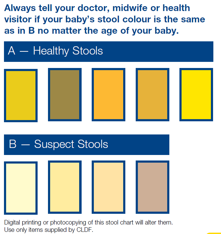
Presents with prolonged jaundice. Dark urine, pale (white!) stools distinguish it from common, benign breast feeding jaundice, but often missed. Parental reporting of stool/urine colour is unreliable! Stool colour chart available from Children’s Liver Disease Foundation.

Normally distal but 20% proximal.

Associated with SPLENIC MALFORMATION syndrome (poly or asplenia, situs inversus, malrotation, absent IVC).

Lanarkshire incidence 1.26 per 10 000, significantly higher than rest of Scotland!  NO evidence of genetic factors.  Pigweed in pregnant ewes in Australia – “biliatresone” toxin. Industrial waste…?

Investigations

Colour of stool!

Fasting (4hrs) USS essential, but sensitivity is operator dependent

Treat by Kasai Porto-enterostomy before 6 weeks ideally (16% normal LFTs, 18% portal hypertension, 94% survival @5yr +/- transplant), else liver transplant.

1yr phenobarb, urso, Vit K.  Long term Dalivit.

Consider varicella vaccination if likely for transplant!

Prognosis

Prognosis related to clearing of jaundice, established cirrhosis/fibrosis, cholangitis, biliary stricture, portal hypertension (degree of – most have).

60% clear jaundice, up to 60% require transplant in first 2yrs.  Of the rest, half need transplant in childhood, leaving just 20% getting to transition with native liver.Most mortality due to transplant complications.

New Japanese data suggests length of jaundice more important than age (traditionally 45-60 days low risk for liver failure)

Outcomes from Kasai operation are better in centres doing more than 5/yr, so only 3 supra-regional centres in England.  But outcomes in Scotland seem to have got worse, even though overall better!  Up to surgeon whether feasible or not for an individual patient.

Increased sepsis due to gut organisms from Roux-en-Y loop.

Cholangitis – features can be seen on USS.  Characteristically unwell, febrile with rise in bilirubin and LFTs (but not always).  Rx Tazocin.  Some require antibiotic prophylaxis.

Portal hypertension can develop early or late.  May present with variceal bleeding, low platelets, splenomegaly. Managed by banding of varices, TIPS shunt, transplant.

Strictures present with biliary stasis, itch, pain, coagulopathy. 

[Rachel Tayler]

SARS

Severe acute respiratory syndrome – caused by one of the coronavirus group, see also MERS and COVID19. The virus probably originated in bats (which commonly carry coronaviruses), then crossed into humans via masked palm civets at the live animal market in Guangdong, China.

The virus spread beyond its original outbreak in China when a businessman became unwell on his flight out of China and died in Vietnam in 2003. Further outbreaks appeared rapidly, as far afield as Toronto. Eventually led to 8000 cases globally, but rapid surveillance and isolation measured brought the epidemic to an abrupt end within 4 months.

Super shedders exist, who have much higher infectivity (1 case on a plane infected 120 others, whereas another plane had 4 cases on board, but no secondary cases occurred!). On the other hand, there is no documented transmission by asymptomatic cases, or between children.

Incubation period is 5-7 but up to 14 days. Spread is by respiratory, fomites, and faecal-oral routes. Peak shedding occurs at peak of clinical disease hence outbreaks were often among health care workers.

Symptoms are ‘flu-like, and non-specific. Fever is universal. Those who do badly have sudden deterioration on 10th day, with ARDS. Mortality is around 10%, but very age dependent, reaching over 50% in the over 65s. Children have lower viral loads, and generally have a benign course. Compared with adults, they perhaps get more gastrointestinal symptoms than respiratory.

Children under 5 yrs are hardly affected at all – perhaps because recent coronavirus infection protective, perhaps because of reduced immune reactivity.

No long term morbidity seen in children.

The diagnosis is suggested by the paucity of clinical signs (mild crepitations only, if anything) with an abnormal chest radiograph (non-specific), and laboratory evidence of leucopenia, lymphopenia, and thrombocytopenia. Raised AST/ALT also seen.

Definitive diagnosis is by ELISA or PCR, neither of which is very sensitive, or useful early on in disease.

Interferon alpha appears to be of benefit in vitro. Otherwise supportive.

Personal Protective Equipment effective if used properly – so buddy system.

Infection control – encourage self isolation, dedicated staff etc.

MERS

Middle East respiratory syndrome, caused by a coronavirus
(MERS-CoV) . See also COVID19 and SARS.

Reported 2012.  More than 2000 cases so far, mostly related to Arabian peninsula, but a single case of MERS-CoV in a returning traveller led to an outbreak involving 186 cases across 16 hospitals in the Republic of Korea.

36% mortality, mostly people with co-morbidities. More than 2000 cases so far. 

One of WHO blueprint priority diseases – potential for serious outbreak, no treatment or vaccine (6-7 others: SARS, Crimean-Congo HF, Ebola, Lassa etc).

Incubation time 2-5 days but up to 14. Median onset to hospitalisation 4 days.

Risk factor appears to be camel contact – milk, meat, urine.

Management

Management based on experience of SARS etc.

Infection control – negative pressure, dedicated staff, cleaning, PPE for suspected cases, self isolation for close contacts. 

COVID19

Hogmanay 2019, WHO were informed of cluster of cases of pneumonia of unknown cause in Wuhan city, Hubei province, China.

Novel coronavirus identified, named SARS-CoV-2. “COVID19” is associated disease. 75% genetically identical to SARS (severe acute respiratory syndrome) and 50% to MERS (Middle East respiratory syndrome) but of course these are both similarly capable of causing severe disease, whereas many coronaviruses pretty benign.

Most likely origin is from live animal markets in Wuhan, although intermediate animal (SARS was found eventually to have crossed over via civet cars). Evidence suggests that there were 2 different llineages in Wuhan, so presumably 2 different Patient Zeroes (which goes against lab leak theory).

By end of February 2020, more than 70 000 cases reported across China, 2500 fatalities. Pandemic was declared by WHO on 11th March.

Cruise ships including the Diamond Princess in Japan (over 700 cases) and the Zaandaam were particularly hard hit.

Lockdown declared in UK on 23rd March 2020.

5 variants of concern, most recently Omicron.

Risk factors

Spike (s) protein binds to ACE2 receptors, primary role of which is to convert AntiThrombin-II into AT-1,7, controlling heart rate, hypertension, vasoconstriction, sodium retention, oxidative stress, inflammation, and fibrosis, as well as enhancing baroreceptor sensitivity. ACE2 variability across populations potentially explaining particular susceptibility among people with hypertension and Africans (nearly double rate of whites) and Asians (although Indian rates lower than Bangladeshi/Pakistani). Rates among Chinese females actually lower than among Whites! [UK data]

At least 3% of severely affected people have known or previously unrecognised genetic defects in type 1 interferon production (especially TLR3 and IRF7 which amplify production).

Risk of “critical illness “ from COVID-19 RR 1.44 if overweight, 1.97 if obese. UK OpenSAFELY analysis. Death 1.27 if BMI 30-39, 2.27 if BMI>40. ACE-2 higher in obese. Plus different immune responses and challenges to ventilate.

London has double the age standardised mortality of any other part of the UK (Birmingham next), as high as 144 per 100 000 in Newham. Glasgow’s rate is about 80 [UK data].

Diabetes, cancer and poorly controlled asthma associated with death in primary care records study. Residential care homes, health care workers, social deprivation, Black/Asian groups also seem to be particularly at risk of death.

Bronx worse hit than Manhattan, despite similar population density. Higher attack and death rates among Afro-Americans. Role for air pollution too?

Plot of mortality rates by gender/race

Pregnancy increases risk slightly, not much risk to baby although elective preterm delivery may be part of management of sick mother.

Acute neurological presentations in adults, including stroke and Guillain Barre syndrome. Thrombosis risk.

Transmission from asymptomatic cases seems to be less important than symptomatic and pre-symptomatic (1-2 days).

In adults, low lymphocytes, high neutrophils and D-dimer predict mortality.

See Treatment.

COVID in Children

Probably more severe than SARS but still children tend to be less severely affected than adults. Cross protection from immunity from other coronaviruses? Differences in ACE2? Some asymptomatic.

16% of hospitalised children admitted to critical care. Age under 1 yr, or age 10-14 yrs, co-morbidities, black ethnicity are risk factors for critical care admission. Mortality rate less than 1% in hospitalised [Swann, ISARIC study]. 3 PIMS deaths in England, all 10-14yrs. 70% of all COVID related deaths in non-white groups. 24% of deaths had no co-morbidities, 60% had life limiting condition. No deaths in kids with asthma, diabetes, Trisomy 21.

Wheeze uncommon.

X-ray more often negative; CT more sensitive.

Can present with GI symptoms.

One baby born to an infected mother developed severe complications.

Neutrophil and LDH counts go up, lymphocytes go down.

A small series of children with COVID-19 has shown a greater prevalence of peripheral halo (halo-sign) lung consolidations on CT.

The criteria for the definition of Acute Respiratory Distress Syndrome (ARDS) and septic shock, the guidelines for the management of sepsis and septic shock and the use of non-invasive ventilation in children are different from those of adults.

Children desaturate more easily during intubation; therefore, it is important to pre-oxygenate with 100% O2 with a mask with a reservoir before intubating.

A rectal swab may be useful in children to determine the timing of the termination of quarantine.

[Chengdu and Italian experience, from PIPSQC]

WHO supports use of dexamethasone in patients with acute respiratory presentation and hypoxia (sats<90%), tachypnoea, or severe respiratory distress. RECOVERY trial continues to study dexamethasone in neonates, plus roles for azithromycin and toculizimab.

Sotrovimab is first line treatment, Remdesivir second line is licensed in hospitalised patients in oxygen, over 12 years and over 40kg and can be considered in this age group for patients with high-risk comorbidity for non-hospitalised patients also. Treatment should be commenced within 5 days of symptom onset (Sotrovimab), within 7 days of symptom onset (remdesivir). Paxlovid (Nirmatrelvir plus Ritonavir) is alternative first line option in adults.

Paediatric multi inflammatory syndrome associated with COVID19 (PIMS-TS)

See PIMS.