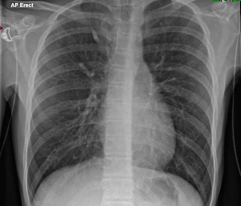
Start outside, work in – soft tissues, then bones, then lungs/heart, finally neck/infradiaphragmatic.
Safety check – position of lines/tunes, check apices for pneumothorax, any foreign bodies?
Adequacy
Rotation – look at symmetry of clavicles and anterior rib ends.
If clavicles high, then lordotic film. May obscure apices.
Penetration – should just be able to make out intravertebral spaces, without lung fields being too dark.
Inspiration – hila become artificially prominent if underinflated.
Thymus
Pesky thing! Can look like pneumonia. Latter more likely if air bronchograms, volume loss (displaced fissure/trachea/mediastinum), effusion. Classically:
indentations where ribs overlie.
Pointy outside edge (“sail sign”).
No mass effect
Lowish density – should still be able to see vascular markings of lung behind
Spinnaker sign is where pneumomediastinum around thymus creates long curving line.
Other normal things
Azygos lobe – normal variant where RUL has near vertical line curving up and out, from thick point (anomalous azygous vein) – giving impression of mediastinal mass.
Azygos lobe at upper right
Mach effect – a line parallel to heart border, looks like pneumocardium but actually optical illusion where your eye “detects” border where there isn’t one…
One diaphragm usually higher than other – both ok, as long as no more than 2cm (one rib space).
Other
Hilum – rings or tram lines suggest bronchitis. Round opacity adjacent to and larger than ring suggests vascular prominence due to left to right shunt.
Silhouette sign – where heart border and/or diaphragm obscured in lower zone due to consolidation in lower lobe (left or right).
Effusion – vertical line at costophrenic angle.
Round pneumonia – will have air bronchograms, compare mass.
Collapse vs consolidation – sharp lower border is the fissure so if deviated then collapse.
Pneumothorax – lucency without clear edge may suggest lung hyperinflation eg bronchial atresia.
If edge projects below diaphragm then likely to be skin fold!
Foreign body – get expiratory film, which will enhance air trapping.
Diagnosis is about probability – high probability is recurrent episodes of cough, wheeze, breathlessness, chest tightness plus documented wheeze, atopic history, documented variable PEF or FEV1. Isolated episodic cough is not sufficient. Episodes typically triggered by viral infections, cold air, exertion, laughter or emotion. Start treatment, “typically” 6 weeks inhaled corticosteroids (ICS). If good response to treatment, then diagnosis is confirmed.
If intermediate probability then spirometry with reversibility is preferred initial test for children old enough to do it (Grade D recommendation). If spirometry normal, then do challenge tests and/or Fractional exhaled nitric oxide (FeNO) measurement. For younger children, watchful waiting or trial of treatment [colour code suggests this is appropriate from age 1, but no advice given for under 1…].
FeNO has reasonable positive predictive value, but false positives in allergic rhinitis, rhinovirus and dietary nitrates, plus overlap in values between asthmatics and normal population (especially children).
Red flags –
Focal chest signs
Abnormal voice or cry
Failure to thrive
Vomiting
Wet/productive cough
Nasal polyps
Management
Self management education, written personalized plan. Assess control – consider using Asthma Control Test (ACT) questionnaire or similar.
Assess risk of future attacks. Co-morbid atopic conditions, younger age, obesity, and exposure to environmental tobacco smoke are markers of increased risk (some of these strongly socioeconomically linked, of course).
Ask specifically about medication use and assess prescriptions. Explore attitudes to medication as well as practical barriers to adherence.
Not for routine house dust mite avoidance measures. Avoid smoking and second hand smoke.
Weight loss (including dietary and exercise programmes) for overweight and obese. Breathing exercise programmes can be offered as an adjuvant to pharmacological treatment for adults.
Treatment
ICS are recommended preventer. An asthma attack in the previous 2 years, symptoms 3 days a week, or using reliever 3 days a week, or waking 1 night a week are indications. Give twice daily at least until good control established.
Start at dose appropriate for the severity of the disease. In mild to moderate asthma, no benefit in starting at high dose and weaning. In children, “reasonable” starting dose is Very Low (100mcg twice daily of Clenil or equivalent).
5yrs and over, if add-on is required then choice between inhaled long acting beta agonist (LABA) or leukotriene receptor antagonist (LTRA). Only then increase dose of ICS from very low (100mcg Clenil or equivalent twice daily) to low (200mcg twice daily).
For exercise induced symptoms, generally just a sign that inadequate control! But if otherwise well controlled then give inhaled short acting beta agonist immediately prior to exercise. Then choice between LRTA, LABA, cromoglicate or theophylline.
Acute Severe Asthma
Sats under 92%
PEF 33-50% of best or predicted
Can’t complete sentences in one breath, or too breathless to feed
HR >140 (under 5), >125 (over 5)
RR>40 (under 5), >30 (over 5)
Life threatening defined as:
PEF <33%
Exhaustion, poor resp effort [tautology?]
Hypotension
Cyanosis
Silent chest
Confusion
Treat –
Oxygen
MDI plus spacer if mild/moderate
If refractory to beta agonist, add ipratropium 250mcg mixed into beta agonist [same dose for everyone]
“Consider adding 150mg magnesium sulphate to each neb in first hour if symptoms started <6hrs and presenting with sats <92%” = 0.3ml of 50% MgSO4
Give oral steroids early, dose by age.
Second line treatment –
Consider single IV bolus of salbutamol (15mcg/kg over 10mins). For bolus dilute to 50mcg/ml with saline/glucose. For infusion, dilute to 200mcg/ml
Consider aminophylline for severe asthma unresponsive to maximal doses of bronchodilators and steroids. Loading dose slow injection over 20 mins! Then dilute to 1mg/ml with saline
Consider IV MgSO4 40mg/kg over 20 mins – dilute to 10% in saline or glucose.
Systematic review of IV Magnesium in children (2018) – pulmonary function improved, hospitalization and further treatment decreased. MAGNETIC trial of Magnesium nebs did not show a clinically significant improvement in mean asthma severity scores in children with acute severe asthma. But better Asthma Severity Score at 1 hour where saturations <92% at presentation and those with preceding symptoms lasting less than 6 hours [Lancet 2013]. 2022 Metanalysis found no benefit but varying protocols and populations.
According to the 2010 Global Disease Burden Assessment, outdoor air pollution caused more than three percent of the annual disability and life lost. Rising due to urbanisation. Responsible for 50 000 deaths annually in the UK.
Air pollution associated with low birth weight, smaller heads, developmental disorders eg autism, type 2 DM, strokes, heart attacks (atherosclerosis), cognitive decline, slower development of lung function with reduced adult capacity (implication for COPD), onset of asthma, wheeze. Not just exacerbations of chronic lung disease!
Different kinds of pollution – particulates (different sizes eg PM1), nitrogen dioxide, sulphur dioxide. Most PM10 from traffic, but natural sources too eg pollen, soil. Wood burners! NO2 and SO2 falling as fewer power stations and less industrial output, but NO2 particular problem for urban centres where most commercial vehicles run on diesel.
Diesel engines also produce polycyclic aromatic hydrocarbons eg BaP (Benzo pyrene), maternal exposure a concern as linked to mental health and neurodevelopmental problems in children. Some also carcinogenic.
Particulates a problem for respiratory conditions. Often contain spores and pollen. Ozone associated with airway hyperresponsiveness.
Not just about degree of pollution – metereological factors (temperature, atmospheric pressure, low humidity etc) complicate. In Taiwan, pollution synergistic with dust mites for development of asthma.
Carbon deposits found in fetal side placental macrophages.
MRSA and stenotrophomonas colonization in CF associated with maternal PM levels.
European study of 325 000 adults found mortality increased proportionally with levels of particulate matter, nitrogen dioxide and black carbon – even at levels below current EU/US/WHO standards. [BMJ 2021;374]
Southern California reduced PM levels and found less severe chronic lung problems.
1 hour commuting in Sao Paolo estimated to be equivalent to 5 cigs/d. In London, travel to school is bulk of exposure (plus school breaks! Note locations!) esp stationary traffic.
What cars produce in lab tests is not the same as in the real world, even when manufacturers don’t cheat!
Low emission zones generally exclude cars, and may just divert traffic elsewhere, not much evidence that they help. London low emission zone has reduced NO2 slightly only. Plan for ultra low zone.
The virus spread beyond its original outbreak in China when a businessman became unwell on his flight out of China and died in Vietnam in 2003. Further outbreaks appeared rapidly, as far afield as Toronto. Eventually led to 8000 cases globally, but rapid surveillance and isolation measured brought the epidemic to an abrupt end within 4 months.
Super shedders exist, who have much higher infectivity (1 case on a plane infected 120 others, whereas another plane had 4 cases on board, but no secondary cases occurred!). On the other hand, there is no documented transmission by asymptomatic cases, or between children.
Incubation period is 5-7 but up to 14 days. Spread is by respiratory, fomites, and faecal-oral routes. Peak shedding occurs at peak of clinical disease hence outbreaks were often among health care workers.
Symptoms are ‘flu-like, and non-specific. Fever is universal. Those who do badly have sudden deterioration on 10th day, with ARDS. Mortality is around 10%, but very age dependent, reaching over 50% in the over 65s. Children have lower viral loads, and generally have a benign course. Compared with adults, they perhaps get more gastrointestinal symptoms than respiratory.
Children under 5 yrs are hardly affected at all – perhaps because recent coronavirus infection protective, perhaps because of reduced immune reactivity.
No long term morbidity seen in children.
The diagnosis is suggested by the paucity of clinical signs (mild crepitations only, if anything) with an abnormal chest radiograph (non-specific), and laboratory evidence of leucopenia, lymphopenia, and thrombocytopenia. Raised AST/ALT also seen.
Definitive diagnosis is by ELISA or PCR, neither of which is very sensitive, or useful early on in disease.
Interferon alpha appears to be of benefit in vitro. Otherwise supportive.
Personal Protective Equipment effective if used properly – so buddy system.
Infection control – encourage self isolation, dedicated staff etc.
Middle East respiratory syndrome, caused by a coronavirus (MERS-CoV) . See also COVID19 and SARS.
Reported 2012. More than 2000 cases so far, mostly related to Arabian peninsula, but a single case of MERS-CoV in a returning traveller led to an outbreak involving 186 cases across 16 hospitals in the Republic of Korea.
36% mortality, mostly people with co-morbidities. More than 2000 cases so far.
One of WHO blueprint priority diseases – potential for serious outbreak, no treatment or vaccine (6-7 others: SARS, Crimean-Congo HF, Ebola, Lassa etc).
Incubation time 2-5 days but up to 14. Median onset to hospitalisation 4 days.
Risk factor appears to be camel contact – milk, meat, urine.
Management
Management based on experience of SARS etc.
Infection control – negative pressure, dedicated staff, cleaning, PPE for suspected cases, self isolation for close contacts.
Hogmanay 2019, WHO were informed of cluster of cases of pneumonia of unknown cause in Wuhan city, Hubei province, China.
Novel coronavirus identified, named SARS-CoV-2. “COVID19” is associated disease. 75% genetically identical to SARS (severe acute respiratory syndrome) and 50% to MERS (Middle East respiratory syndrome) but of course these are both similarly capable of causing severe disease, whereas many coronaviruses pretty benign.
Most likely origin is from live animal markets in Wuhan, although intermediate animal (SARS was found eventually to have crossed over via civet cars). Evidence suggests that there were 2 different llineages in Wuhan, so presumably 2 different Patient Zeroes (which goes against lab leak theory).
By end of February 2020, more than 70 000 cases reported across China, 2500 fatalities. Pandemic was declared by WHO on 11th March.
Cruise ships including the Diamond Princess in Japan (over 700 cases) and the Zaandaam were particularly hard hit.
Lockdown declared in UK on 23rd March 2020.
5 variants of concern, most recently Omicron.
Risk factors
Spike (s) protein binds to ACE2 receptors, primary role of which is to convert AntiThrombin-II into AT-1,7, controlling heart rate, hypertension, vasoconstriction, sodium retention, oxidative stress, inflammation, and fibrosis, as well as enhancing baroreceptor sensitivity. ACE2 variability across populations potentially explaining particular susceptibility among people with hypertension and Africans (nearly double rate of whites) and Asians (although Indian rates lower than Bangladeshi/Pakistani). Rates among Chinese females actually lower than among Whites! [UK data]
At least 3% of severely affected people have known or previously unrecognised genetic defects in type 1 interferon production (especially TLR3 and IRF7 which amplify production).
Risk of “critical illness “ from COVID-19 RR 1.44 if overweight, 1.97 if obese. UK OpenSAFELY analysis. Death 1.27 if BMI 30-39, 2.27 if BMI>40. ACE-2 higher in obese. Plus different immune responses and challenges to ventilate.
London has double the age standardised mortality of any other part of the UK (Birmingham next), as high as 144 per 100 000 in Newham. Glasgow’s rate is about 80 [UK data].
Diabetes, cancer and poorly controlled asthma associated with death in primary care records study. Residential care homes, health care workers, social deprivation, Black/Asian groups also seem to be particularly at risk of death.
Bronx worse hit than Manhattan, despite similar population density. Higher attack and death rates among Afro-Americans. Role for air pollution too?
Pregnancy increases risk slightly, not much risk to baby although elective preterm delivery may be part of management of sick mother.
Acute neurological presentations in adults, including stroke and Guillain Barre syndrome. Thrombosis risk.
Transmission from asymptomatic cases seems to be less important than symptomatic and pre-symptomatic (1-2 days).
In adults, low lymphocytes, high neutrophils and D-dimer predict mortality.
Probably more severe than SARS but still children tend to be less severely affected than adults. Cross protection from immunity from other coronaviruses? Differences in ACE2? Some asymptomatic.
16% of hospitalised children admitted to critical care. Age under 1 yr, or age 10-14 yrs, co-morbidities, black ethnicity are risk factors for critical care admission. Mortality rate less than 1% in hospitalised [Swann, ISARIC study]. 3 PIMS deaths in England, all 10-14yrs. 70% of all COVID related deaths in non-white groups. 24% of deaths had no co-morbidities, 60% had life limiting condition. No deaths in kids with asthma, diabetes, Trisomy 21.
Wheeze uncommon.
X-ray more often negative; CT more sensitive.
Can present with GI symptoms.
One baby born to an infected mother developed severe complications.
Neutrophil and LDH counts go up, lymphocytes go down.
A small series of children with COVID-19 has shown a greater prevalence of peripheral halo (halo-sign) lung consolidations on CT.
The criteria for the definition of Acute Respiratory Distress Syndrome (ARDS) and septic shock, the guidelines for the management of sepsis and septic shock and the use of non-invasive ventilation in children are different from those of adults.
Children desaturate more easily during intubation; therefore, it is important to pre-oxygenate with 100% O2 with a mask with a reservoir before intubating.
A rectal swab may be useful in children to determine the timing of the termination of quarantine.
[Chengdu and Italian experience, from PIPSQC]
WHO supports use of dexamethasone in patients with acute respiratory presentation and hypoxia (sats<90%), tachypnoea, or severe respiratory distress. RECOVERY trial continues to study dexamethasone in neonates, plus roles for azithromycin and toculizimab.
Sotrovimab is first line treatment, Remdesivir second line is licensed in hospitalised patients in oxygen, over 12 years and over 40kg and can be considered in this age group for patients with high-risk comorbidity for non-hospitalised patients also. Treatment should be commenced within 5 days of symptom onset (Sotrovimab), within 7 days of symptom onset (remdesivir). Paxlovid (Nirmatrelvir plus Ritonavir) is alternative first line option in adults.
Paediatric multi inflammatory syndrome associated with COVID19 (PIMS-TS)
The most common inherited genetic condition in N European populations, with carrier rate of 1 in 20. Incidence is therefore around 1 in 4000 births. Most common gene defect is deletion at delta F508 of CFTR (CF transmembrane conductance regulator) gene.
Features:
family history
congenital intestinal atresia
meconium ileus
distal intestinal obstruction syndrome
faltering growth (in infants and young children)
undernutrition
recurrent and chronic pulmonary disease, such as:
recurrent lower respiratory tract infections
clinical or radiological evidence of lung disease (in particular bronchiectasis)
persistent chest X-ray changes
chronic wet or productive cough
chronic sinus disease
obstructive azoospermia (in young people and adults)
acute or chronic pancreatitis
malabsorption
rectal prolapse (in children)
pseudo-Bartter syndrome.
Median predicted life expectancy is now nearly 50 years, but this doesn’t take into account the new CFTR modulators. Management has evolved slowly, with revolutionary improvements including high calorie diets/feeding, pancreatic enzyme replacement, specialist CF centres and CF newborn screening.
Drugs
Regular prophylactic antibiotics (usually starting with oral flucloxacillin) introduced at early stage. Later on may require regular courses of IV antibiotics.
Azithromycin given on Mondays, Wednesdays and Fridays – but for anti-inflammatory properties as much as antibacterial.
Creon is the name for pancreatic enzyme supplements, taken with each meal.
Fat soluble vitamins.
Nebulised DNAse and hypertonic saline to help with chest physiotherapy
New treatments
CFTR modulators treat the basic defect. Lead to significantly improved pulmonary function, decreased respiratory infections and improved nutrition.
Combination elexacaftor, tezacaftor and ivacaftor, will be suitable for approximately 90% of all people with CF. But expensive, with many countries unwilling or unable to fund them.
CFTR phenotypes vary. Class I–III variants are most severe, with minimal or no CFTR function. Class IV–VI variants are where CFTR is produced and reaches apical membrane but doesn’t work normally, so milder phenotype.
Ivacaftor reduces hospital admission, rates of respiratory Pseudomonas and Aspergillus infection, and halves rate of decline in FEV1 %, suggesting at least 5 years survival benefit.
For suspected asthma, where child unable to do spirometry, then watchful waiting or trial of treatment for specified time period. Choice of treatment depends on severity and frequency of symptoms – “typically 6 weeks inhaled steroid”, “very low dose”.
Start regular preventer treatment or escalate treatment if you are getting frequent symptoms, viz:
three times a week or more, or
using your blue inhaler three times a week or more, or
if your asthma is waking you up once a week or more.
Start regular preventer if asthma attack in previous 2 years!
Same table for all ages now, and same steroid doses!
Step 1 – very low dose inhaled corticosteroid (ICS). OR leukotriene receptor antagonist (LRTA) if under 5.
Step 2 – Add LRTA if under 5, else inhaled long acting Beta agonist (LABA) if 5+.
Step 3 – If no response to LABA, stop and increase ICS dose. If some benefit from LABA continue and increase ICS dose, or consider trial of LTRA.
Step 4 – high dose therapies: increase ICS dose to medium, or add slow release theophylline. Refer for specialist care.
ICS doses
Very low dose is 50mcg 2 puffs twice daily of beclometasone. Low dose is double that, medium 200mcg 2 puffs twice daily.
QVAR and fluticasone are double the efficacy of beclometasone so doses are halved. Ciclesonide is somewhere in between.
Incubation period 1-4 days. Infectious period from day before symptoms appear, to 5 days after symptoms appear. Virus shedding can persist for months in immunocompromised (as other viruses).
Main types A and B. B shows antigenic drift, with minor variations over time. A shows antigenic shift, with appearance of new N type usually associated with global pandemic eg Spanish flu. Influenza viruses circulate through birds and pigs, so new types typically occur through reassortment of genes across different species (Chinese food markets are a perfect petri dish).
Case definition
At least one of these systemic symptoms: Fever (or feverishness), malaise, headache, myalgia;
PLUS at least one of these respiratory symptoms: cough, sore throat (!), shortness of breath.
Transmission
Large droplet eg sneezing, range only a metre or so.
Direct/indirect contact eg sneezing into hands, and then to surfaces. Survive at least 24 hrs in environment.
Aerosol generating procedures can produce small droplets with further range (how far?). Includes intubation, extubation, open suctioning, CPAP/HFOV/BiPAP, CPR. NOT high flow O2 or nebs.
Prevention is therefore by good hygiene viz covering nose and mouth during coughing/sneezing, wiping with disposable tissues, avoiding touching nose/eyes/mouth, washing hands (alcohol gel adequate if hands visibly clean); and immunisation.
For close patient contact, aprons, masks, gloves recommended. Similarly, consider eye protection.
For aerosol generating procedures, FFP3 (filter face piece, efficiency grade 3) masks required. Such masks can be worn for up to 8 hours if necessary. Fluid repellent gowns if extensive secretions or bodily fluids anticipated.
Complications
Pneumonia, ARDS
Meningitis/encephalitis – see below
Myositis and rhabdomyolysis leading to kidney failure (esp flu B)
Reyes syndrome (liver failure and encephalitis, also associated with aspirin)
Encephalitis lethargica? Probably not… Epidemic around time of Spanish influenza pandemic, fever then neuropsychiatric deterioration – estimated 1m cases globally, high mortality. Cf Subacute sclerosing panencephalitis (measles).
Encephalitis
Neurological presentations are mostly transient, improving within 48 hours. Pre-existing neurological disorders more at risk (unusually severe seizures in known epileptic children, mostly).
Rarely acute necrotizing encephalitis (treat with high dose steroids). CT can show (classically bilateral thalamic necrosis) but MRI better of course. Not just influenza but viral, and immune mediated (no virus found in brain/CSF). Familial susceptibility described due to RANBP1 mutation. [2024 Review from Toulouse]
Vaccination
See immunisation. Antigenic drift means new influenza vaccines need to be developed each year, reflecting the common serotypes affecting people in other parts of the world who have already had their winter.
2021 metanalysis (37 studies) found immunisation in children was 53.3% effective against hospitalization (68.7% vs flu A/H1N1pdm09 specifically).
Only 44.3% for live-attenuated influenza vaccines cf 68.9% for inactivated vaccines.
Sphingomyelinase (lysosome) disorder. Type B has only visceral involvement, can survive into adulthood. More common in Ashkenazi Jews.
Typically develop symptoms at around 6 months. Can be prolonged jaundice as baby, else abdominal distension (hepatosplenomegaly), growth failure, hypotonia, failure to meet milestones.
Death usually around 3yrs, recurrent lung infections, interstitial lung disease. Spasticity develops later.
Cherry red spot seen on fundoscopy in macula, possibly not in early stages.