Category Archives: Genetics

Neurofibromatosis

Autosomal dominant condition, so 50% risk of passing on to offspring and variable penetrance. May be spontaneous mutation.

Diagnosis still essentially clinical: 1988 Consensus is for any 2 of the following:

  • 6 or more cafe-au-lait spots (at least “pencil thickness” in diameter?)
  • axillary or groin freckling
  • 2 or more Lisch nodules – pigmented lesions in iris, may need slit lamp to see. No clinical consequence!
  • 2 or more neurofibromas,
  • optic pathway glioma (OPG),
  • bone dysplasia,
  • first-degree family relative with NF1 

Plexiform neurofibromas are larger ones with potential to cause visual, hearing, even cardiac problems.

Issues are:

  • High blood pressure
  • Epilepsy (lesions develop in brain)
  • Bone problems including scoliosis
  • Effects of neurofibromas in sensitive locations
  • Cosmetic appearance, self image
  • Cancer (malignant change in neurofibroma but also brain and breast)
  • Risk to own children

Ophthalmology review annually in children, every 2 yrs in adults, given risk of optic gliomas.

Family support at https://nervetumours.org.uk/

Clinical trial of selumetinib had beneficial effect on inoperable plexiform fibromas.
G6PD deficiency

Glucose 6 phosphate dehydrogenase deficiency. X-linked.

Can present with prolonged jaundice in babies. Otherwise with haemolysis (causing jaundice and anaemia).

Haemolysis triggered by infections, but also drugs and chemicals. Classically sulfonamides, but most relevant are:

  • Anti-malarials (ironically)
  • Nitrofurantoin!
  • Aspirin (so Kawasaki)
  • Henna! Other dyes
  • Moth balls!

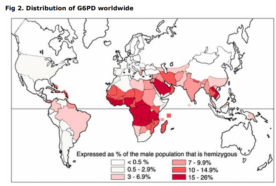
Geographical risk areas –

Porphyria

Various types, all accumulate toxic levels of porphyrins in body but variable presentations.

Variegate Porphyria

The one associated with Dutch or Afrikaner (white South African) genetics. PPOX mutations

Specific symptoms can vary greatly; autosomal dominant so variable penetrance (can even be asymptomatic). Typically:

  • Photosensitivity
  • GI symptoms including abdominal pain, nausea, vomiting, constipation
  • Neurological: extremity pain, weakness, anxiety, restlessness and convulsions
  • Acute attacks, usually severe abdominal pain (can be chest), vomiting first. Then confusion, convulsions, and muscular weakness. Lasts days or weeks. Recovery from severe paralysis can be slow.

Triggers for acute attacks include drugs, hormones (especially progesterone), lack of carbohydrate intake, alcohol, and stress.

Diagnosis is by porphyrins in plasma, urine and stool.

Treatment now available – Panhematin is used to halt acute attacks and prevent attacks, Givlaari just for prevention.

Congenital cataracts

Differential:

  • Varicella
  • Rubella
  • CMV
  • Toxoplasmosis
  • HSV
  • Syphilis, HIV
  • Genetic non syndromic – various
  • Syndromes – Downs/Patau
  • X-linked Lowe syndrome (LD and proximal tubular dysfunction)
  • Autosomal dominant myotonic dystrophy (various eye problems), Neurofibromatosis type 2
  • Zellweger (includes neonatal adrenoleukodystrophy and infantile Refsum) – peroxisomal
  • Galactosaemia, Cockayne syndrome

Heterotaxy syndromes

A spectrum of disorders where the normal left-right arrangement of organs in the body is disturbed (“errors of lateralisation”).

Situs inversus is complete mirror image arrangement – there are no health consequences as a result (other than iatrogenic eg delayed diagnosis of appendicitis).

Many genetic causes. Primary cilial dyskinesia (Kartagener syndrome) is one.

Heart – IVC can be interrupted, requiring azygos veins to drain lower body vessels back to heart. Many variations of valves and connections seen. Congenital heart block often seen.

Gut – malrotation, biliary atresia

Asplenia or polysplenia.

Horseshoe or dysplastic kidneys.

Cowden syndrome

Autosomal dominant, PTEN gene (10q23). See OMIM.

Clinically –

  • Macrocephaly
  • Skin lesions – esp hamartomatous. Eg trichilemmomas (smooth, skin coloured, warty or dome like lesions, esp face), acral keratoses (ie on hands), papillomatous papules)
  • Increased risk for the development of breast, thyroid, and endometrial carcinoma

In some cases intestinal polyps, papilloedema, immunodeficiency.

Bartter’s syndrome

Abnormal renal excretion, leading to low potassium.

Presents in early childhood with failure to thrive. Could also be constipation, muscle cramps and weakness (potassium needed for membrane potential, so these are all neuromuscular) and non-specific dizziness and fatigue.

Characteristic hypokalemic, hypochloremic metabolic alkalosis. High plasma renin activity and high aldosterone concentration seen.

Gitelman syndrome is similar, less severe (distal tubule, rather than ascending limb of loop of Henle) – less failure to thrive, in fact often asymptomatic detected incidentally. Might present with nocturia/polyuria.

Urinary calcium excretion distinguishes the two syndromes. Bartter’s waste calcium (more severe, after all), Gitelman retain.

Treatment is with supplementation.

Decompensation can be precipitated by diarrhoea or vomiting. Acute treatment can include potassium-sparing diuretics (spironolactone), cyclo-oxygenase inhibitors and renin-angiotensin blockers.

Pseudo-Bartter’s is due to CF.

Fragile X

Cause of developmental delay.

FMR1 gene is on X chromosome, obviously, and is a trinucleotide repeat disorder (along with Friedrich’s ataxia, myotonic dystrophy, Huntington disease etc), so inheritance is interesting.

Dads can carry gene, but only pass it on to their daughters (who will all get it).

Mums will carry gene on 1 chromosome, so sons and daughters can both get it, but 50:50 chance.

As with other trinucleotide repeat disorders, gene expands with each generation, so risk of disease increases from 1 generation to the next, and this is somewhat predictable: intermediate gene (so 45-54 copies) won’t expand to cause disease (200+ copies) in 1 generation, but premutation gene (55-199) copies probably will.

Features:

  • Moderately severe learning disability
  • Facial features – long face, midface hypoplasia, large lips and jaw, small ears
  • Macro-orchidism

Females less severely affected, of course.

Rhabdomyolysis

Muscle breakdown with release of products into blood stream that can cause acute renal failure. Can be associated with compartment syndrome, disseminated intravascular coagulation.

Typically occurs with crush injuries, sometimes seen with extreme endurance sports.

Can be infectious.

Recurrent seen with fatty acid oxidation disorders and Lipin 1 mutations.

Lipin 1 mutations

Autosomal recessive – heterozygotes may have exercise induced muscle symptoms or be prone to drug induced myopathy.

Basal CK high but spikes to over 100 000 with decompensation (infection, exercise, anaesthetic).

Treat crises with carbohydrates/intralipid.

Muckle-Wells Syndrome

A Cryopyrin disorder, found in Northern Europeans. Cryopyrin triggers an IL-1 dominated inflammatory response, and is coded for by the Cold-Induced Autoinflammatory Syndrome 1 (CIAS1) gene, also known as the NLRP3, NALP3 or PYPAF1 gene.

Attacks of periodic fever are very brief eg 1-2 days – apart from fever, an urticarial rash is sometimes seen, limb pain/arthralgia occurs. Abdominal pain and arthritis occur rarely. Sensorineural hearing loss is characteristic.

Amyloidosis affects 25%, which is high cf other periodic syndromes.

Diagnosis is by genetics.

Steroids are often used but benefit is inconsistent; interleukin 1 (IL-1) receptor antagonist Anakinra shows promise.

Familial cold autoinflammatory syndrome (FCAS) is a similar condition, also related to Cryopyrins. Cold induced obviously, but without the deafness, and amyloidosis is rare.

NOMID/CINCA are also related – the names say it all: Neonatal onset multisystem inflammatory disorder, and chronic infantile neurological cutaneous and articular syndrome. Papilloedema and uveitis potentially leading to blindness occur; there is epiphyseal bone formation; hepatosplenomegaly; and a chronic meningitis with deafness. There is no known treatment, sadly.