Category Archives: Neonatology

Biliary atresia

Wasting of biliary tree +/- gall bladder in early months of life (LANDING’s theory). Premature babies get it less (as wasting hasn’t progressed as much) but can still get it!

Stool colour chart

Presents with prolonged jaundice. Dark urine, pale (white!) stools distinguish it from common, benign breast feeding jaundice, but often missed. Parental reporting of stool/urine colour is unreliable! Stool colour chart available from Children’s Liver Disease Foundation.

Normally distal but 20% proximal.

Associated with SPLENIC MALFORMATION syndrome (poly or asplenia, situs inversus, malrotation, absent IVC).

Lanarkshire incidence 1.26 per 10 000, significantly higher than rest of Scotland!  NO evidence of genetic factors.  Pigweed in pregnant ewes in Australia – “biliatresone” toxin. Industrial waste…?

Investigations

Colour of stool!

Fasting (4hrs) USS essential, but sensitivity is operator dependent

Treat by Kasai Porto-enterostomy before 6 weeks ideally (16% normal LFTs, 18% portal hypertension, 94% survival @5yr +/- transplant), else liver transplant.

1yr phenobarb, urso, Vit K.  Long term Dalivit.

Consider varicella vaccination if likely for transplant!

Prognosis

Prognosis related to clearing of jaundice, established cirrhosis/fibrosis, cholangitis, biliary stricture, portal hypertension (degree of – most have).

60% clear jaundice, up to 60% require transplant in first 2yrs.  Of the rest, half need transplant in childhood, leaving just 20% getting to transition with native liver.Most mortality due to transplant complications.

New Japanese data suggests length of jaundice more important than age (traditionally 45-60 days low risk for liver failure)

Outcomes from Kasai operation are better in centres doing more than 5/yr, so only 3 supra-regional centres in England.  But outcomes in Scotland seem to have got worse, even though overall better!  Up to surgeon whether feasible or not for an individual patient.

Increased sepsis due to gut organisms from Roux-en-Y loop.

Cholangitis – features can be seen on USS.  Characteristically unwell, febrile with rise in bilirubin and LFTs (but not always).  Rx Tazocin.  Some require antibiotic prophylaxis.

Portal hypertension can develop early or late.  May present with variceal bleeding, low platelets, splenomegaly. Managed by banding of varices, TIPS shunt, transplant.

Strictures present with biliary stasis, itch, pain, coagulopathy. 

[Rachel Tayler]

Alagille Syndrome

Autosomal dominant condition (70% sporadic) with characteristic facies, biliary hypoplasia, vertebral and cardiac abnormalities. JAG1 gene.

Paucity of intrahepatic bile ducts so typically prolonged jaundice, but depending on how many ducts, may not be obvious in first few months. Jaundice gradually improves, but only minority clear completely. 30% progress to cirrhosis.

Posterior embryotoxon (white ring at periphery of cornea) is a clue, but seen in 10% of normal population so not specific.

Cardiac includes pulmonary stenosis, Tetralogy of Fallot.

Facies: broad forehead, triangular face, deepset eyes, long nose with bulbous tip.

Butterfly vertebrae characteristic (asymptomatic – see on chest x-ray).

Other:

  • Renal Tubular Acidosis, renal cysts
  • Growth failure
  • Pancreatic insufficiency

Urate

Product of amino acid metabolism.

In developmental delay, an abnormally high or low result is significant viz:

  • Glycogen Storage disorder
  • Purine disorders eg Lesch Nyhan
  • Molybdenum Cofactor deficiency – other features are microcephaly, hyperekplexia

High urate levels can also be risk factor for urolithiasis (stones in urinary tract)

Niemann Pick disease

Sphingomyelinase (lysosome) disorder.  Type B has only visceral involvement, can survive into adulthood.  More common in Ashkenazi Jews.

Typically develop symptoms at around 6 months.  Can be prolonged jaundice as baby, else abdominal distension (hepatosplenomegaly), growth failure, hypotonia, failure to meet milestones.

Death usually around 3yrs, recurrent lung infections, interstitial lung disease.  Spasticity develops later.

Cherry red spot seen on fundoscopy in macula, possibly not in early stages.

Congenital Syphilis

In US primary, secondary and cong syphilis all surged in the 90s, now focal outbreaks in urban, drug using population. In N Africa, 3% of pregnancies, up to 7% in Carribean. 1 million pregnancies affected worldwide, of which 50% will end in abortion or still birth, and the other 50% will be congenital cases (unlike TORCH).

Clinically, 2/3 affected neonates asymptomatic at birth. Otherwise:

  • “snuffles” (vesicles on upper lip, highly infectious)
  • mucous patches (moist erosions)
  • hepatosplenomegaly and hepatitis
  • anaemia +/- hydrops
  • meningitis, with CSF pleocytosis and high protein
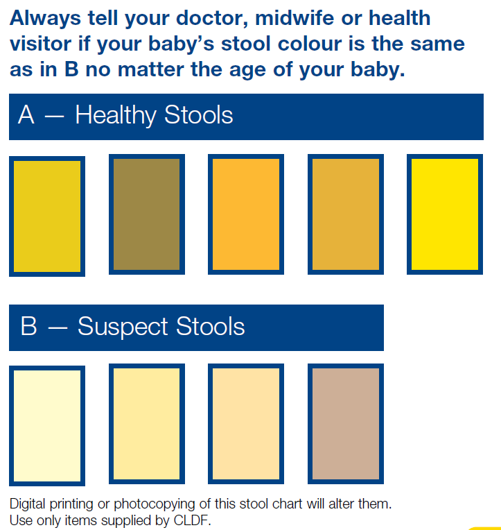
  • pneumonia, +/or fluffy diffuse infitrates on CXR (=pneumonia alba)
  • pseudoparalysis

Years later:

  • Typical facies – frontal bossing, saddle nose, short maxilla, high palate
  • Mulberry Molar (5 blobs in ring shape to tooth, pathognomic!). Hutchinson’s incisors (peg-like) better known.
  • Gummata (rubbery ulcers)
  • Sabre tibia (anterior bowing)
  • Hutchinson’s triad = interstitial keratitis, peg shaped incisors, and sensorineural deafness.

Prevention

The risk of transmission is very high, particularly for untreated primary (ie a chancre) or secondary (ie multiple lesions, lymphadenopathy) disease. The risk falls to 40% for early latent syphilis (ie test positive but asymptomatic, with infection likely to have occurred in the previous 12 months on the basis of previous tests, symptoms or exposure). A category exists of latent syphilis of unknown duration, which only applies to patients aged 13-35yrs with nontreponemal titre of 32 or more.

Treating syphilis in pregnancy – for early acquired disease (primary, secondary or latent of <1yr) benzathine penicillin 50 000U/kg, with second dose a week later if in third trimester [BNF] but exclude neuro. For late latent syphilis >1yr duration give 3 doses at weekly intervals. For neuro disease benzylpen 50 000U/kg qds for 10-14/7 followed by 3 doses benzathine penicillin as above. If HIV positive also, then there tends to be more CNS disease, treatment failure, and treatment reactions (fever, myalgia, preterm labour – give steroids).

Adequate antenatal treatment = adequate benzathinepenicillin dose (2.4M Units IM) once weekly x3 – erythromycin is not reliable), 30 days before birth, proven 4x drop in nontreponemal serology.

Diagnosis

Syphilis tests are either nontreponemal or treponemal.

  • Nontreponemal viz VDRL, RPR are screening tests, 70% sensitive in primary, 99% in secondary.
    • False positives – lupus, infection, recent immunization, pregnancy, other treponemes.
    • Quantitative – correlate with disease activity: 4x rise in titre early on or in relapse, drop of 4x suggests adequate response to treatment. In secondary, titres are always high ie 1:32.
    • False negative – early? Tertiary – V high levels of antibody! So if high suspicion then do dilutions.
    • Should become negative within 1 yr of treatment in primary, 2 yrs in secondary or congenital, 5 yrs in late.
  • Specific treponemal tests eg TP immobilization (TBI), fluorescent T antibody absorption (FTA-Abs) used to confirm.
    • False positive with non pallidum, Lyme or other borrelia.
    • Remain positive for life, even with treatment.
    • Do not correlate with disease activity.
    • FTA-Abs IgM available for testing baby, but still has false positives/negatives

In newborns, direct microscopy and fluorescent antibody tests can be done from mucous patch, else from placenta (beware non pallidum treponemes in normal flora esp mouth). PCR can be done from lesions too. VDRL more than 2x dilutions richer than mum’s is suggestive. IgM can be negative early esp infection occurring late in pregnancy, not always recommended. False positive VDRL may occur due to transplacental antibodies if high maternal levels.

Where disease is likely, or maternal treatment has been inadequate, further testing is required:

  • FBC, LFTs
  • Lumbar puncture, incl VDRL on CSF (not 100% sensitive so if congenital disease suspected, treat for neuro.)
  • XR long bones to look for destructive lesions. Even in asymptomatic, XR changes seen in 20% esp ankles, knees but also wrists, elbows. Lesions are symmetric, multiple: periostitis, osteitis, osteochondritis.
  • CXR
  • Ophthalmology assessment

For screening adults, 1 step strip test available, and one off oral treatment (Azithro, 1.8g). Antibodies give only partial protection.

Treatment

For congenital syphilis treat with benzylpen 100-150 000 U/kg/d in bd or tds doses for 10-14/7.

There is concern about CSF levels with procaine or benzathine penicillin, although sometimes these are used for asymptomatic babies with nontreponemal tests less than 4x mum’s, which might occur with inadequate treatment. In this situation, any abnormal finding on evaluation requires full 10/7 course.

If late diagnosis (>4/52), give high dose viz 200-300 000 U/kg/d qds for 10-14/7. If CNS disease is excluded, this could be converted to benzathine penicillin.

Follow up

Monitor to show fall in VDRL at 3-6 months.

(Rana Chakraborty, St George’s)

Antenatal corticosteroids

Proven to reduce the incidence of respiratory distress syndrome (RDS) due to surfactant deficiency.

Meta-analysis shows benefit even for babies near term ie 34-39/40!  Risk ratio 0.74.

Not much data on long term outcomes, of course, but RDS rates high even at 38 weeks so LSCS should not be planned until 39 weeks.

Repeat courses of antenatal steroids (due to threatened delivery) associated with lower birth weight, so should ideally be limited to 3. [https://journals.plos.org/plosmedicine/article?id=10.1371/journal.pmed.1002771]

Sacral dimple

Typical sacral dimples are <5mm in diameter, within 25mm of anus and located in midline.  Rate of spinal  dysraphism (bifida occulta) less than 1%.

Higher risk if do not fulfill these criteria. Lipomas, deviated/bifurcated crease are the most likely to be associated with dysraphism.  Otherwise you expect at least 2 or more cutaneous markers (hair tuft, haemangioma, Mongolian spot, skin tag/tail).

Reports of high frequency of hair tufts in diastematomyelia probably refer to more striking lesions (“faun tails”).

Royal College of Radiology has policy  – ignore sacral dimples unless atypical, or in combination with other lesions.

USS if neonate, but MRI if US abnormal or equivocal, where neurological signs (bladder, bowel, lower limb) or lesion discharging.

[Arch Derm 2004]

Sudden Unexpected Postnatal Collapse

Identified as a group by BPSU, led by Julie-Clare Becher in Edinburgh.  As with SUDI, previously well appearing baby suddenly suffers cardiorespiratory failure, with death or severe neurological disability as a result.

Her 2011 report highlighted an association with prone position and breast feeding.  One third of of cases had an underlying pathological or clinical condition but over half probably due to accidental suffocation, frequently unrecognised by parents themselves.

Some suggestion that cases are underreported, and that cases are increasing due to increased early skin-skin practices without adequate supervision.  In a big survey from Sweden, most happen within first 2 hours of birth, often during first breast feeding attempt.

The process of referral to procurator fiscal, bereavement support, prolonged delay in post mortem report etc is common to SUDI, even if only a few of the underlying risk factors are shared.

A BAPM protocol is available, detailing elements of history eg family tree, drug/alcohol/smoking history, obstetric history; plus investigations eg Kleihauer, placental histology, skeletal survey, PHOX2B gene (congenital central hypoventilation syndrome), fibroblast culture from skin biopsy.  In a death, most if not all should be done routinely by pathology.

In terms of prevention, standard SUDI precautions are emphasized – avoid prone position, co-bedding, head covering, beware sedating drugs etc.  Education of parents regarding warning signs and upper airway control.  Better non-obtrusive monitoring.

[Arch Dis Child Fetal Neonatal Ed 2012;97:F30−F34. doi:10.1136/F30 adc.2010.208736; Translational Stroke Research , Volume 4, Issue 2, pp 236-247]


Beckwith Wiedemann Syndrome

Chromosome 11p problem, where IGF2 gene lies. Hemihypertrophy, macroglossia, ear creases/pits, exomphalos, umbilical hernia, visceromegaly, macrosomy, hyperinsulinism.

Focal disease is associated with uniparental disomy in chromosome 11 (“upd(11)pat”), whereas diffuse disease can be familial or sporadic.

Focal disease can be managed by limited surgery, whereas diffuse disease needs near total pancreatectomy for any benefit. DOPA-PET scans can differentiate focal disease with a sensitivity of 88-94%, and are 100% accurate in localizing the focal lesion (Aberdeen does).

At increased risk of tumours esp nephroblastoma, adrenal carcinoma, hepatoblastoma. Screening recommendations depend on mutation viz IC2 LOM, IC1 GOM, upd(11)pat etc. For all but the first, every 3 months until age 7yrs.

OMIM link.

Support group https://www.bwssupport.com/

Congenital Hyperinsulinism

Hyperinsulinaemic hypoglycaemia is a common cause of persistent severe hypoglycaemia, and is associated with long term neurodisability and epilepsy. Can be congenital, due to unregulated beta cell function (used to be called nesidioblastosis), else secondary to various other conditions eg:

  • Maternal diabetes mellitus
  • Birth asphyxia
  • IUGR
  • Beckwith Wiedemann Syndrome – about 50% are affected, usually transiently
  • Inborn error of metabolism eg glycosylation disorder

Can present with severe hypoglycaemia in the newborn period despite good oral intakes, else more insidiously in infancy and childhood. No ketones of course – cf ketotic hypoglycaemia.  Macrosomia (+/- hypertrophic cardiomyopathy and hepatomegaly) is a feature of fetal hyperinsulinism but is not always present, otherwise typical features of hypoglycaemia may be seen, including seizures.

Cases secondary to IUGR/asphyxia tend to be transient, settling within a few days, but some have persistent symptoms for months before suddenly resolving!

Recessive defects in sulphonylurea receptor probably most common cause.  Fasting or exertion are the usual triggers, but postprandial symptoms may reflect dumping syndrome (usually secondary to gastro-oesophageal surgery) or else hyperammonaemic hyperinsulinism (glutamate dehydrogenase disorder – the high ammonia of 100-200 is persistent but asymptomatic). A form of exercise induced hyperinsulinism exists that requires exercise testing to diagnose.

Insulinoma is more likely in later childhood. Can be part of a multiple endocrine neoplasia syndrome type 1 (ask family history).

Raised plasma hydroxybutyrylcarnitine and urinary 3-hydroxyglutarate are diagnostic of Hydroxy AcylCoA Dehydrogenase (HADH) deficiency.

Management aims to achieve normal glucose levels, using Carbohydrate supplemented oral feeds plus IV fluids. A central line may be needed to give concentrated dextrose solutions, esp if chubby. Glucagon can be given IM in an emergency but may lead to paradoxical insulin release. Glucagon and/or octreotide can be given as infusions in resistant cases.

For ongoing treatment, diazoxide acts on K-ATP channels in beta cells. Chlorthiazide is synergistic in the neonatal period. Hypertrichosis is the main side effect of diazoxide. Some rare gene defects are resistant to Diazoxide! Feeding probs esp GORD common, partly due to all the NG/IV feeding.

[Kapoor, Arch Dis Child 2009 PMID 19193661]