Category Archives: General paediatrics

Chronic Granulomatous Disease

A neutrophil defect, prediposes to certain characteristic organisms. Frustrated neutrophils cause granulomas, hence the name: these are responsible for some other characteristic features of the disease.

Due to NADPH oxidase mutations, causing reduced oxidative burst (needed by neutrophils to kill pathogens after phagocytosis). Various types depending on which subunit affected. X linked gp91phox def (ie NADPH OXidase, due to CYBB gene defect) is severe, p47phox is autosomal and milder (NCF1 gene, partial activity retained). Other gene defects are CYBA and NCF2, funnily enough, and RAC2. Phenotype varies even between mono twins (factors?). Since non-oxidative killing is then the only remaining immune mechanism, the most important pathogens are catalase positive (an enzyme to make bugs resistant to the peroxide and superoxide produced by neutrophils or by the bacteria themselves as a toxic byproduct). Register exists in UK.

Usually present by 2 yrs but occ not until adulthood. Most commonly lymphadenitis (often culture negative), skin infections (esp perianal), pneumonia. Hepatomegaly is a clue. Other sites of infection are osteomyelitis, liver abscesses (distinctive, fibrous capsule, septated, thick contents – vague presentation, blood cultures usually negative so low index of suspicion).

Probably not the superoxides themselves that are important; MPO deficiency does not tend to present with infections! Critical step is probably degranulation of primary granules, with release of elastases etc. Superoxides probably deal with toxic metabolites rather than doing the killing.

Chronic inflammatory problems occur (due to having intact upregulated phagocytic activity with reduced apoptosis):

  • BCG causes lymphadenitis in CGD, but does not disseminate.
  • colitis (characterized by pigment laden macrophages, quite specific for CGD but not very sensitive). Seen in 45% of X-linked, 10% of autosomal recessive. Tends to present with abdo pain rather than frank bleeding, often subclinical, may account for FTT.
  • lupus like rash and illness
  • Poor wound healing (classically dehiscence at 10/7 post op, non-purulent)
  • cystitis
  • pericarditis
  • chorioretinitis
  • Hollow organs may become obstructed by granulomas esp oesophagus, gastric outlet, bladder.
  • pulmonary fibrosis

Mums who are carriers of CGD can be symptomatic even with fairly decent percentages of functional neutrophils incl aspergillus! Due to lyonization.  Onset variable.

Diagnosis

Nitroblue tetrazolium (NBT) test now replaced by Dihydrorhodamine test on flow cytometer (false positive esp in preterm so always check with reference lab). Only takes 15 mins!

The organisms found are also characteristic:

  • Staphylococcus aureus – found in most liver abscesses as well as in skin
  • Enterobacteria – Enterobacter, E coli, Salmonella, Klebsiella, Aerobacter, Serratia, Yersinia, Proteus
  • Aspergillus – mostly fumigatus but A. nidulans is emerging in US, and Candida albicans, Scedosporium apiospernum and Chyrosporium zonatum reported. Can be acute, esp after inhalation eg digging in garden. Biopsy may be needed to make diagnosis. Steroids useful for severe inflammatory disease!!!
  • Burkholderia cepacia – looks identical to Pseudomonas, so any Ps not specified as aeruginosa should be considered suspect! (Pseudomonas itself is catalase positive but susceptible to non-oxidative killing so is not a problem).
  • Septicaemia is v rare ! – but may be seen with B. cepacia.
  • Nocardia common in US, rare in Europe – gram positive soil organism, forms filaments like a fungus. Usually pneumonia but also skin, CNS. Sensitive to co-trimoxazole.
  • Actinomycosis – even though catalase negative!

Note that you do NOT see PCP, strep, onychomycosis (despite susceptibility to fungus) or lymphomas (cf granulomas).

Patients are often anaemic with an iron-deficient pattern but resistant to iron supplementation (except in bowel disease, ?vitamin B12 def). ESR is often raised even when well. Less of a problem with CRP.

Management

Co-trimoxazole prophylaxis (daily dosing, not 3x weekly as in PCP prophylaxis) good because active against typical bugs, and intracellular. Itraconazole is prophylactic vs Aspergillus.

Avoid BCG because of tendency to form abscess.

Cipro and Fluclox good for first line – effective against typical organisms, and cipro acts intracellularly. In serious pneumonia, empirical treatment should consist of Ceftazidime/Meropenem, Fluclox and Amphotericin. Because of the range of possible organisms, bacteriological diagnosis should not be delayed and tissue biopsy sought if non-invasive methods unsuccessful.

Steroids for colitis, cystitis and obstructive granulomatous disease. Also for poor wound healing!

Other adjuncts:

  • Voriconazole is an effective oral antifungal, so useful when no tissue diagnosis (beware accumulation of amphotericin with long term use, causing permanent renal damage).
  • Interferon (IFN) gamma – effective as prophylaxis in large Multicentre study, but strong centre effect with less benefit in Europe (in fact the lowest incidence seen with antibiotic prophylaxis) so not universally used (but safe). Less evidence in established infection but that’s when it tends to get used! Increases NO production by neutrophils, by improved RNA splicing efficiency(?). Give three times a week by subcut injection; side effect is fever and flu-like symptoms. Better antifungals mean it is used less now.
  • Granulocyte infusions (apheresed from donors after GCSF priming) can be done every 1-2 days (or instilled directly into lesions) but rarely needed now with better antifungals. Increases risk of reaction to Ambisome, plus you become sensitized, which will hinder transplant prospects. Can support infected kids through BMT.
  • Surgery may be necessary to remove infected tissue, may help get positive culture.
  • BMT is indicated at diagnosis if a matched donor is available.
  • Gene therapy has produced transient improvements only.

Median survival 30 years – NB compliance with prophylaxis by adolescents. So consider transplant.

[Clin Exp Imm 122(1); October 2000 pp 1-9 GOLDBLATT, D; THRASHER, AJ]
Recurrent boils

Potentially symptom of diabetes, chronic granulomatous disease, Hyper IgE syndrome – but more usually just an individual thing, or a nasty strain of staphlycoccus aureus.

GOS says if otherwise well (no other abscesses, no colitis, no weird organisms eg Serratia) then consider eradication with:

  • Naseptin – a medication taken four times a day for a period of 10 day.s
  • Chlorhexidine shower or bath and hairwash every day for a period of 14 days.
  • Keep a separate towel for each member of the family, change for a clean towel every two days and wash the dirty towels on a hot wash cycle.

[http://www.gosh.nhs.uk/medical-information-0/search-medical-conditions/recurrent-boils]

Fatty Acid Oxidation Disorders

Various eg CoA disorders eg MCAD, LCAD, VLCAD; Carnitine disorders (transports fatty acids into mitochondria). Present with severe hypoglycaemia.

There are related lipid storage disorders eg Fabry, Niemann Pick, MCLD where hypoglycaemia is not a feature.

AST/ALT raised, due to protein breakdown for gluconeogenesis. Acylcarnitines, organic acids abnormal.

MCAD

=Medium Chain Acyl CoA Dehydrogenase deficiency. Can be asymptomatic eg parents of newly diagnosed child, even with same gene defect! Crisis – vomiting, hypoglycaemia, hyperammonaemia, sudden death.

Diagnosis: Octanoyl- acylcarnitine increased.

Management is by avoidance of fasting , plus carnitine! Newborn screening started in UK in 2009.  

Carnitine deficiency

In primary deficiency, there is non ketotic hypoglycaemia and cardiomyopathy, hepatomegaly, hyperammonaemia.

Various other abnormalities. Usually acylcarnitine, organic and amino acid analysis will clarify.

Glycogen Storage Disorders

Various. Not a problem of storing it, a problem of breaking it down! Classic type 1 is Glucose -6-phosphatase deficiency. Depending on the type, gluconeogenesis as well as glycogenolysis may be impaired – some of the enzymes are involved in both – so hypoglycaemia with ketones, lactate and triglycerides high. Liver becomes enlarged with excessive glycogen, Glucagon has no effect.

Managed by regular meals and extra complex carbohydrate eg cornstarch, as for ketotic hypoglycaemia.

Glycogen synthase deficiency is sometimes included. If you can’t make glycogen then you get an immediate glucose dip post prandially, you don’t get a big liver (obviously) but other mechanisms work ok so lactate is normal (cf typical Glycogen storage disorder).

Pompe disease is a lysosomal disorder, infantile form affects heart, neurodevelopment (enzyme treatment available).

McArdle syndrome is myophosphorylase defect – pain/weakness/cramps on exertion, myoglobinuria, second wind phenomenon (rapid recovery with rest).

Ketotic hypoglycaemia

What happens when you don’t eat enough and your carbohydrate stores run out!  Typically due to illness, especially with vomiting.

But  can be endocrine cause eg hypopituitarism, adrenal insufficiency. Growth hormone deficiency associated with recurrent hypoglycaemia even before growth failure apparent, associated with sudden death.

If you have excluded glycogen storage disorders (big liver, high lactate), glycogen synthase deficiency (normal liver, high lactate) and organic acidurias (acidosis, encephalopathy, usually high ammonia too), can be idiopathic (usually SGA at birth, thin, presents under 4yr, resolves by 7yr).

If ketones low and fatty acids high, then suggests fatty acid oxidation disorder (but usually just means hypoglycaemia was treated before sample was collected).

Regular meals + night time complex carbo snack, optimize nutrition, carbs if unwell eg Maxijul + Electrolade else Ribena/apple juice.

Childhood absence epilepsy

Age 3 to 10 yr, girls more than boys

Abrupt onset and cessation

Stare into space, unresponsive for around 10-20 seconds

Afterwards immediately carry on unaware anything has happened – or possibly very brief confusion (seconds)

Clue that not just day dreaming is that most also have clonic movements, minor changes in tone (eg head drops, or held object dropped), automatisms (repetitive movements of eyes, mouth).

No myoclonus (else likely to be juvenile myoclonic epilepsy). Some get generalised tonic-clonic seizures in adolescence but these are infrequent and respond well to treatment.

Usually when sat quietly rather than actively engaged in something. Precipitated by sleep deprivation, as other epilepsies, but also hyperventilation (90% detected after 3 minutes – get them to count aloud)

Atypical would be gradual onset, last longer, have more obvious changes in tone – but continuum

Genetics

Most genetic. GLUT1 (glucose transporter protein type 1, coded for by SLC2A1 gene, involved in glucose transfer across blood-brain barrier) mutations are present in up to 10% of early childhood absence epilepsy (ie under 5yrs). Heterozygous mutations are mostly de novo but may be inherited as AD trait. SLC2A1 positive often not helped by valproate and ethosuximide, whereas ketogenic diet often effective.

EEG

EEG shows sudden onset 3Hz spike and wave, esp with photic stimulation/hyperventilation. Interictal is often normal. Clinically apparent if more than 3 seconds of activity, but detailed neuropsych assessment suggests that non-clinical absences do cause functional impairment. Atypical have slower spike waves and rarely have normal interictal.

Prognosis

60-80% full remission, usually during puberty; in most cases, absences disappear on monotherapy but there are resistant cases (unpredictable, other than SLC2A mutations).

Occipital lobe epilepsy

Early (PANAYIOTOPOULOS) or BECOP (Benign epilepsy of childhood with occipital paroxysms).

Panayiotopoulos

  • Or Self limiting epilepsy with autonomic symptoms (SeLEAS)
  • Usually 3-5yrs but can be 1 to 15
  • Mostly nocturnal
  • Characteristically autonomic – pallor/flushing/vomiting/coughing!
  • Head/eye deviation, then tonic-clonic (one sided or generalized)
  • Often status, but actually seizures infrequent
  • EEG can be normal – sleep EEG more sensitive
  • Good prognosis – most remit with a few years and have just a few seizures
  • Valproate or carbamazepine

BECOP

  • Usually middle childhood, cf above
  • Can be triggered by going from dark to light areas or vice versa
  • Visual  – partial or complete loss, flashing lights, multi-coloured circles, balls, rarely hallucinations
  • Headache during or after – cf migraine!
  • Often one side jerks; rarely generalized
  • Carbamazepine or Valproate
  • Usually benign, most remit by puberty

Urine collection

For culture, looking for urine infection, traditionally midstream urine, or at least clean catch.  Hard if not toilet trained.

Ideally, avoid first portion of sample (easier if toilet trained), more likely to be contaminated.

Need to be well hydrated, of course.

In/out catheter and suprapubic aspiration are quick but invasive and unpleasant.

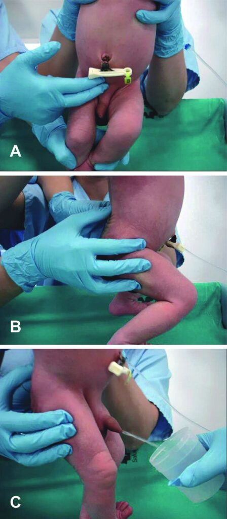
For babies, “Dangle-tap” urine collection method –

  • Feed first!
  • Then hold the baby under their armpits with their legs dangling (parent can do). Another person then starts bladder stimulation – gentle tapping in the suprapubic area at a frequency of 100 per minute for 30 s.
  • The third step is stimulation of the lumbar paravertebral zone in the lower back with a light circular massage for 30 s. Repeat as necessary.
  • Babies hate the suprapubic tapping, but often pee when you switch to back rub.

86% successful within 5 minutes, mean 57secs! Over 3 months hard as heavy and actively resisting.

Older babies – try Quick Wee – gentle suprapubic cutaneous stimulation using gauze soaked in cold fluid for 5 mins. 30% successful within 5 minutes.

[Madrid Infanta Sofia hospital, as reported in Arch Dis Child
2013;98:27-29 doi:10.1136/archdischild-2012-301872]]

Non-specific effects of childhood vaccines

Systematic review.

BCG and Measles vaccine appear to reduce all cause mortality, not just TB and Measles (relative risk 0.7 in trials, 0.5 in observational studies, roughly same for both vaccines).

That’s wonderful, until you see that DTP vaccine appears to increase mortality (in 7 of 9 observational studies).

So are live vaccines “immune boosting” in some general way, whereas inactivated vaccines are not, or actually deleterious?

Large proportion of data from studies in Guinea-Bissau so high risk of bias!

[BMJ 2016; 355]

Systematic review of immunological effects of BCG found evidence of changes in non-specific immunological variables (eg IFN-gamma prodution, enhanced lymphocyte proliferation in response to candida, HBsAg, staph etc) but inconsistent, dubious clinical relevance, and certainly not geographically generalisable.

Ultimately, better designed studies, linked to epidemiology, needed before policy changes can be justified.