Category Archives: Cardiology

Alagille Syndrome

Autosomal dominant condition (70% sporadic) with characteristic facies, biliary hypoplasia, vertebral and cardiac abnormalities. JAG1 gene.

Paucity of intrahepatic bile ducts so typically prolonged jaundice, but depending on how many ducts, may not be obvious in first few months. Jaundice gradually improves, but only minority clear completely. 30% progress to cirrhosis.

Posterior embryotoxon (white ring at periphery of cornea) is a clue, but seen in 10% of normal population so not specific.

Cardiac includes pulmonary stenosis, Tetralogy of Fallot.

Facies: broad forehead, triangular face, deepset eyes, long nose with bulbous tip.

Butterfly vertebrae characteristic (asymptomatic – see on chest x-ray).

Other:

  • Renal Tubular Acidosis, renal cysts
  • Growth failure
  • Pancreatic insufficiency

Murmurs

An added sound heard when listening with a stethoscope, distinct from heart sounds or other clicks or snaps.

Can indicate a structural abnormality.  But can be heard in normal hearts too, esp kids.

Still’s murmur

Or “innocent” murmur.  Characteristic vibratory, low pitched, crescendo-decrescendo sound – “like an Aeolian harp” – loudest along left sternal border.  Never louder than grade 3.  Typically gets quieter when child sits or stands up (reduced venous return?) – you would not expect a murmur caused by a structural abnormality to change much.

Present at any age. Usually goes away by adolescence.

Venous hum

Another innocent one, a rumble heard in the upper chest, disappears when lying down, or when neck turned or neck veins occluded gently.

Pulmonary flow murmur

Pulmonary valve closest to anterior chest wall, which might explain why you sometimes hear this.  Might be confused with pulmonary stenosis or subaortic membrane.

Prolonged QT interval

An abnormal finding on ECG.

QT interval changes with heart rate, so usually calculated as corrected QT (QTc), where average QT is divided by square root of RR interval (ie 1 second, if heart rate 60).

Associated with dysrhythmia, especially torsades de pointes (polymorphic ventricular tachycardia).

Seen with:

  • Genetic predisposition – Long QT syndrome
  • Certain drugs – antipsychotics (eg chlorpromazine, quetiapine), antiarrhythmics (!?), tricyclic antidepressants (eg amitriptyline), other antidepressants (eg citalopram, venlafaxine), antihistamines (terfenadine, but also loratadine, diphenhydramine, astemizole), macrolides, quinine.

Cerebral Arteriovenous malformation (AVM)

Present with hydrocephalus or else cardiac failure in neonatal period. Clue is that heart is structurally normal!

Life time risk of haemorrhage is high, presenting with headache or seizure.  Annual rate about 6%.  Focal neuro signs rare prior to haemorrhage!  Risk slightly higher in deep locations eg basal ganglia.

Once haemorrhage has occurred, risk of further haemorrhage increases significantly.  Microsurgery, radiosurgery or embolization techniques used alone or in combination.


Rheumatic fever

Rare in developed world now, still common in underdeveloped world, or at least in underdeveloped communities eg Aboriginal Australians.  Prob also genetic susceptibility.

Caused by Group A streptococcus.  Important cause of acquired heart valve disease.  Can recur.

Probably cross reactivity between specific Group A strep M proteins and human tissues.

Erythema marginatum
Erythema marginatum

Diagnosis

Jones criteria:

  • Major
    • Carditis eg new murmur.  Mitral most commonly, classically apical blowing pan-systolic.  Aortic next most common.
    • Arthritis esp large joints.  Migratory.
    • Subcutaneous nodules – these are the most uncommon major criterium (in Turkish study of over 1000 cases there were none with nodules).  Typically over extensor surfaces of joints, 0.5-2cm, symmetrical.
    • Sydenhams chorea
    • Erythema marginatum – not specific to rheumatic fever. Seen in 0.4% of Turkish study patients. Serpiginous or annual eruption, can look similar to erythema multiforme. Provoked by warmth eg bath.  Non pruritic.
  • Minor
    • Fever
    • Arthralgia
    • Prolonged PR interval on ECG
    • Elevated CRP/ESR
  • 2 major or 1 major plus 2 minor, plus confirmation of group A streptococcal infection eg positive culture, high ASO titre sufficient for diagnosis. Modified Jones takes into account background incidence.

Note that initial infection may be subclinical eg pharyngitis, erysipelas. Symptoms of rheumatic fever develop 10 days to several weeks later. Chorea can appear months later.  Low threshold for echo as carditis can also be subclinical.

Established criteria for rheumatic valvulitis – Gewitz 2015

Treatment

Antibiotics – Treat with penicillin,  this does not however affect clinical course but hopefully prevents further spread of that particular bug. Traditionally single dose intramuscular Penicillin G Benzathine.

NSAIDs for joint pain.  Usually dramatic response, if not then reconsider diagnosis!

Valproate for chorea, possibly steroids – see Sydenham’s.

Aspirin and/or Steroids for carditis, but not much evidence.  Diuretics, ACE inhibitors for cardiac failure.

Long term treatment

Recurrence with progression of valve damage is the main concern, and well recognized. Subclinical carditis improves in about 50% but definite risk of progression (mild definite and borderline RHD showed 26% and 9.8% echocardiographic progression respectively).

Regular intramuscular penicillin (benzathine pencillin G) every 2-3 weeks has the lowest recurrence rates but oral penicillin V more acceptable.  Erythromycin or cephalexin if allergic.

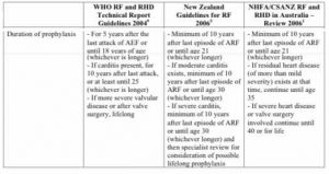
WHO recommendations:

  • Rheumatic fever without carditis: 5 years after last attack or until age 18 (whichever is longer)
  • Rheumatic fever with carditis but without residual disease: 10 years after last attack or until age 25 (whichever is longer)
  • Residual valve disease or valve replacement: lifelong

American and Australian heart association guidelines vary slightly:

Penicillin prophylaxis guidelines comparison

Familial Hypercholesterolaemia

Usually LDL receptor gene mutations (on liver, for removing lipids from blood). One Apo-B mutation (on LDL) identified.  Least common is PCSK9 mutation – higher degradation of receptor, most severe.

If normal BMI, elevated LDL, autosomal dom inheritance, normal TFTs then 95% LDL receptor defect.

If one sibling has a LDL double that of another sibling, 99.8% LDL receptor mutation.

Highly heterogeneous in UK, over a 1000 mutations.

Estimated 1/500 in most countries [but in Denmark found 1/137, Holland has found 1 in 232].  So prob only 12% of cases identified in UK, even less in young individuals!

DNA screening cf lipid testing cost effective (and enhances cascade screening).  Using LDL only, 8-15% false neg and pos, due to overlapping of ranges.  Gene scoring (using SNPs) for mutation neg clinical cases helps to find polygenic cases. But 40% less risk of disease so cascade testing less effective.

American college of med genetics have recommended that familial hypercholesterolaemia (FH) mutations detected on whole genome should be reported to patient.

Cases of sudden cardiac death in 4-6yr olds reported in homozygous.  Heterozygous cases still important – 100X coronary risk age 20-40yrs…

Diagnosis

Diagnosis in childhood could save 50 healthy life years!!!  Dutch cascade screening since 1994 stopped last year. Only 1/3 of known paed cases came clinically, hence big gap in case finding.

Simon Broome criteria – (else use Dutch system) distinguishes Definite FH vs Possible.

  • Definite = DNA pos for known mutation, else tendon xanthomata (personal or 1st or 2nd deg relative) plus cholesterol as below.
  • Possible = cholesterol concentrations as below PLUS:
    • Family history of myocardial infarction: aged younger than 50 years in second-degree relative or aged younger than 60 years in first-degree relative.
    • Family history of raised total cholesterol: greater than 7.5 mmol/l in adult first- or second-degree relative or greater than 6.7 mmol/l in child, brother or sister aged younger than 16 years.

Cholesterol cut offs for Simon Broome

Total cholesterol LDL-C
Child/young person > 6.7 mmol/l > 4.0 mmol/l
Adults > 7.5 mmol/l > 4.9 mmol/l

Refer both definite and possible cases for DNA testing. Plus there are age/sex specific cholesterol cut offs for relative of a known case where DNA is negative.  NICE standard is that children with possible FH should be offered diagnostic tests by age 10.

Given how prevalent use of statins is now, it’s harder to get a positive family history! So history of grandparents more important! 10% have no family history

Only 4% of heterozygotes have xanthomas (can be in buttock cleft, but only look if found elsewhere!). Arcus at 6 oclock in children, all round in adults. Palpable in Achilles tendon rather than seen? Pain in achilles often reported! Resolve with treatment.

Management

NL guidelines – 5-7yr diet etc only.  Diet with plant sterols can drop LDL by 7%

Over 10yrs, treatment (high dose, low dose, combination) depends on mutation and LDL. Most experience with pravastatin.

LDL apheresis as treatment.

Excellent safety and tolerance of statins. Significant reduction within 2 years.

Like any regular medication, compare it to toothbrushing.  Do both at the same time!

Most cardiologists don’t consider FH when high cholesterol found. Nurses? Via labs?

Check growth, puberty, healthy lifestyle. Update family pedigree, contraception.

Smoking confers massive additional risk.  Albert Wiegman has had a 5yr old admitting to smoking…

Treating from age 6.

Outcome

10yr follow up submitted – mean age 24yrs. 7 already older than the age their parent had event. LDL still higher than sibs.


Boys have higher risk of cardiac death then girls, but smoking gives girls worse risk than boys.

Intima media thickness – increases year on year, so earlier treatment the better?  Even before age 8 significantly different from non FH.


In NICE audit, majority of children not on treatment, and being seen in adult lipid clinics.

No evidence that low fat diets etc are affecting growth (limited numbers so far).

Homozygous FH being recommended as rare disease, so deserving of national specialist service.


Long QT Syndrome (LQTS)

16 genes now known, most incomplete penetrance. Important in drug induced Torsades too. LQTS probably accounts for many sudden unexpected deaths, some evidence for SUDI too.  Some genes associated with epilepsy. Triggers for cardiac event seem to vary from 1 gene to another eg exercise (LQT1), sudden loud noise (LQT2).

Variable expressivity too, so 1 normal ECG not adequate.

Other ECG clues are bradycardia, T wave alternans or biphasic, abnormal U waves. T wave changes with posture or exercise may also be seen. Allow at least 3 consecutive beats to measure, to allow for sinus arrhythmia.

Consensus diagnostic criteria

Any of:

  1. LQTS risk score >=3.5 (in the absence of an alternative cause) – score is complex, but involves QTc, Torsades, T wave abnormalities, syncope, cong deafness, FH.
  2. Pathogenic mutation in a LQTS gene
  3. QTc >500 on repeated ECG
  4. QTc 480-499 (repeated) PLUS unexplained syncope.

Treatment is with beta blockers, avoidance of triggers.

[Current Opinion in Pediatrics. 26(6):727-33, 2014 PMID: 25313972].

Lyme Disease

Borrelia (spirochaete) infection, spread by ticks (Ixodes), common in localized areas of Europe and North America (forest environments).

Differential includes possible co-infection from other tick born organisms viz anaplasmosis, or babesiosis.

Vaccine available if likely to be at risk.

Clinical

Infection occurs a minimum of 48 hours after bite!

Skin – Erythema migrans is the classic skin lesion, a spreading ring usually at the site of the bite but can be multiple and at different sites.  Typically not hot, itchy or painful. Takes a while for central clearing to develop. Develops over 1-4 weeks (from 3 days to 3 months!), can last months.  Looks like erythema multiforme, but time scale different.  Insect bite hypersensitivity/superinfection looks similar but usually hot, itchy and/or painful, and develops/recedes within 48 hours!

Lyme lymphocytoma is a painless bluish red nodule or plaque, especially on the ear but also reported on the nipple and scrotum.  More common in children.  May persist for months, can precede other features.  Acrodermatitis chronica atrophicans (ACA) is almost exclusively seen in adults, predominantly women, and is an eruption with chronic, progressive red or bluish-red lesions, usually on the extensor surfaces, with later atrophic, fibroid or sclerodermic changes.

Consider Lyme as possible (but unlikely) cause for:

  • fever and sweats
  • swollen glands
  • malaise
  • fatigue
  • neck pain or stiffness
  • migratory joint or muscle aches and pain
  • cognitive impairment, such as memory problems and difficulty concentrating (sometimes described as ‘brain fog’)
  • headache
  • paraesthesia

Arthritis – uncommon, presents as recurrent inflammation of 1 or more large joints, usually the knee. Swelling can be disproportionate to pain.  Can become more persistent – in a minority, despite treatment, inflammation becomes chronic (presumably immune-mediated).

Carditis occurs rarely, and almost always with other clinical features.  Usually partial heart block, but can be complete, usually resolves within a week.

Neurological – isolated facial palsy, meningitis, other cranial nerve palsies, meningoencophalitis, polyradiculopathy.  There is a small proportion of children who can present with non-specific headache, fatigue, neck pain without clear neurological signs (and also the rare case of raised intracranial pressure).

Other rare disease manifestations include uveitis, iridocyclitis and keratitis.

Diagnosis

For erythema migrans, clinical diagnosis is adequate, and antibodies only positive in 30-70% anyway!

Use a combination of clinical presentation and laboratory testing to guide diagnosis and treatment in people without erythema migrans. Do not rule out diagnosis if tests are negative but there is high clinical suspicion of Lyme disease.

  • Offer an enzyme-linked immunosorbent assay (ELISA) test for Lyme disease – consider starting treatment with antibiotics while waiting for the results if there is a high clinical suspicion. (Test for both IgM and IgG antibodies)
  • If the ELISA is positive or equivocal, perform an immunoblot test for Lyme disease (again, consider starting treatment with antibiotics while waiting for the results if there is a high clinical suspicion). [Western blot increases specificity, but cut offs (for both serology and Western blot) can be an issue, with potential false positives for other acute infections and autoimmune conditions.  Definitely needs to be an approved lab…]
  • If ELISA negative and the person still has symptoms, review their history and symptoms, and think about the possibility of an alternative diagnosis.  If tested within 4 weeks from symptom onset, repeat the ELISA 4 to 6 weeks after the first test.
  • If Lyme disease is still suspected in people with a negative ELISA who have had symptoms for 12 weeks or more, perform an immunoblot test.  If negative, consider synovial fluid aspirate/biopsy, or lumbar puncture [PCR – culture is difficult – or CSF antibodies for neuroborreliosis; consider for isolated facial palsy]
  • If immunoblot negative and symptoms resolved, no treatment is required.

For early neuroborreliosis, antibodies 80% sensitive, rises to virtually 100% for late or ACA.

Early antibiotic treatment is also believed to potentially block antibody production.

Antibodies can then persist for months or even years after successful treatment of the infection, so repeat testing is not useful for monitoring treatment success.

First line ELISA test can have false positives for other spirochaetes, glandular fever and autoimmune conditions.

The idea that there are seronegative “chronic Lyme” cases has little evidence to support it, with only 2 possible cases reported (ACA and arthritis, not neuro).

NICE says “Discuss the diagnosis and management of Lyme disease in children and young people under 18 years with a specialist, unless they have a single erythema migrans lesion and no other symptoms. Choose a specialist appropriate for the child or young person’s symptoms dependent on availability, for example, a paediatrician, paediatric infectious disease specialist or a paediatric neurologist.”

Treatment [check NICE]

The most commonly recommended first-line treatments for different stages of Lyme borreliosis in Europe are:

  • Erythema migrans/borrelial lymphocytoma:  10-14 days Doxycycline if 9yr+ (initially 5 mg/kg in 2 divided doses on day 1, then 2.5 mg/kg daily in 1–2 divided doses, max dose 200mg, for a total of 21 days, option for higher dosing) – 10 days courses of doxy effective in US trials.  Else Amoxicillin 50mg/kg/d, max 500mg TDS (10-14 days)[BNFc says 30mg/kg/d, max 1g, TDS for 21 days].  Don’t delay treatment pending test results.  Scandinavia use 10 days Pen V (100mg/kg/d, max 1000mg TDS). BNFc says Azithromycin as alternative.
  • Isolated facial palsy: 14 days Oral doxycycline  – else as above.  Doesn’t probably help resolution but may prevent later complications.
  • Meningitis/radiculopathy: PO Doxycycline or IV Ceftriaxone  50-100mg/kg/d, max 2g daily (14-21 days). [BNFc talks about CNS disease separate from cranial/peripheral nerves]
  • Encephalitis, myelitis: Ceftriaxone (14 days)
  • Lyme arthritis: Doxycycline (28 days) else Amoxicllin (21-28 days)
  • Carditis: Ceftriaxone during pacing, else PO doxycycline (14 days)

Ceftriaxone is the most commonly preferred parenteral agent, with once-daily dosing facilitating outpatient treatment. Recent prospective studies have shown that oral doxycycline is noninferior to ceftriaxone in neuroborreliosis, and it is now recommended in Europe for the treatment of acute facial palsy (FP), meningitis and radiculoneuritis. Ceftriaxone currently remains the preferred choice for children with other presentations of neuroborreliosis and for those with contraindications to doxycycline.

Several recent EM treatment studies have incorporated noninfected control groups. Excellent responses were seen, with resolution of rash within 7–14 days. Nonspecific symptoms including headache, myalgia, arthralgia, fatigue and parasthesias were no more common in cases than controls at 6-month follow up.

[position statement by the British Infection Association, J Inf 2011;62:329]

[Pediatric Infectious Disease Journal Volume 33(4), April 2014, p 407–409]  

ECG

Standard ECG settings are 25mm/s and 10mm/mV.  Beware small complexes, which may indicate someone has adjusted the Y-axis to 5mm/mV.

Axis @ birth 60-180, @ 1 yr 10-100, @ >1yr 30-90 (NB prems have LESS Rt dominance!). So you expect QRS to be positive in both I and aVF. Under 1, I can be negative but aVF should still be positive.

RVH defined as:

  • Dominant RV1 +/- Q after 1 yr,
  • Upright TV1 over 1 week and under 7-8 yr,
  • SV6 over 15mm if under 1yr or over 5mm if over 1 yr.

Brugada syndrome – genetic arrhythmia. Persistent or intermittent right sided ST elevation and RBBB, leading to VF and sudden death.

QTc is normally under 0.490 up to 6 months, or 0.425 if over 6 months. Calculation = QT/SQR(RR), where QT is time from beginning of Q to end of T. Easiest way to calculate it is to count small squares: QTc is then QT/5 divided by SQR(RR) (where default is 25mm/sec). Over 450ms with history of syncope is a red flag; otherwise over 460ms. Consider family history of sudden death or “epilepsy”, too.

PR 80-160ms (so 3-5 small squares), QRS less than 75ms. Look for delta wave if PR short.

RV1 less than 20-26mm (trough at 1 year), SV6 less than 10mm at birth falling to 4 mm at 10 year.

LVH – deep Q waves in V6 are a clue (upper limit is 0.54mV, ie 5 small squares). The SV1+RV6 upper limit ranges from 3.1mV (newborns) through to 5.7 (older children), staying at around 5 for most children (ie 50 small squares, my calculations). Glasgow guideline says if V5 (rather than V1) and V6 complexes overlap but this depends on how the ECG is printed and can very between 20 and 40 squares!!

For adults, there are many different criteria for LVH eg:

  • Sokolow + Lyon (Am Heart J, 1949;37:161)
    • S V1+ R V5 or V6 > 35 mm
  • Cornell criteria (Circulation, 1987;3: 565-72)
    • SV3 + R avl > 28 mm in men
    • SV3 + R avl > 20 mm in women

But sensitivity of ECG criteria less than 20% at specificity levels of 88% to 92%. Obesity affects chest lead voltage, for example.  Better in patients with a specific cardiac disease. An elevated LVM (left ventricular mass) index is taken as the reference for LVH. In kids the SV3R + RV7 Sokolow-Lyon parameter performs best, but who does V7 routinely? (and still only 25.3% sensitivity).

So you can say voltage criteria for LVH met, but can’t say diagnostic of LVH.  More likely when other features such as left axis deviation, ST and T wave changes. ST elevation under 2 squares not significant. T wave inversion in V5/6 most suspicious (Glasgow guideline). When clinical evidence is also taken into account, the sensitivity improves considerably (but still under 50%).

[Normal ranges – European Heart Journal (2001) 22, 702–711]

LVM (as estimated from echocardiographic measurements) is itself vulnerable to measurement error and may oversimplify the geometry of the left ventricle. Alternatively, a combination of increased LVM and clinical evidence of volume or pressure overload of the left ventricle may be a better reference standard.