Used to work out whether biochemical abnormalities are due to renal dysfunction. There is not really a “normal range” for sodium and potassium in the urine, because it depends whether the body is trying to retain or excrete at any given time. So urinary sodium can be undetectable in dehydration, for instance.
Since creatinine is filtered passively, you can compare how much sodium/potassium is being excreted with what you would expect, by calculating:
Sodium excretion (Urinary Na/Plasma Na), divided by creatinine clearance (urinary creatinine/Plasma creatinine). Multiply by 100 to get a percentage.
If sodium low, you expect the kidneys to retain, so fractional excretion should be less than 1%. For low potassium, fractional excretion should be less than 10%. The opposite is true for high values.
Even where plasma sodium normal, fractional excretion can give you a clue to kidney disease – 1-4% suggests intrinsic renal pathology, over 4% post-renal.
Renal causes of low sodium/potassium include renal tubular acidosis (various forms), Bartter’s syndrome. Non-renal causes include GI losses (eg pyloric stenosis), Pseudo-Bartter’s syndrome (eg CF).
An alternative, possibly simpler method is transtubular potassium gradient (TTKG) :
For this formula to be accurate urine osmolality should be higher than plasma osmolality and urine sodium should be greater than 25 mEq/L.
Individuals with hyperkalemia should have a TTKG above 10. Values below 7 are consistent with mineralcorticoid deficiency, especially if accompanied by hyponatremia and high urine sodium concentration.
Individuals with hypokalemia should have TTKG values below 2.
Can be due to mineral deficiency or toxicity. But can become habitual, in which case motives/consequences should be explored – attention? Escape? Sensory feedback?
Usually iron deficiency, but potentially calcium, zinc. Beware vitamin deficiencies esp C.
Lead exposure can come from toys sourced from outside EU. Houses in area built before 1950? Water companies generally screen for this, houses are occasionally notified of a hazard. But lead poisoning can also be a consequence of pica.
Complications are rare but potential for bezoar formation, gastrointestinal side effects. Toxocariasis if faeces is ingested.
Management
Ignore or avoid negative attention (eye contact, facial expression, speech)
Other oral stimulation eg. chew wristbands
Reward keeping hands in pockets?
Teach edible vs. Non-edible
Alternative communications methods
Provide similar smells, textures, colours to play with or eat
Vast group of vegetables. As with all cross reactivity, allergy is not automatic but being allergic to one or more increases the chance you will be allergic to another.
Disseminated BCG reported, implies SCID or similar major immunodeficiency.
Severe BCG reaction can also indicate underlying TB infection!
More common issues are BCG abscess, and lymphadenitis.
Abscess at injection site appears after a few weeks, can persist for months. Treatment with isoniazid has been offered but no evidence of benefit. Incision probably makes things worse!
Non-suppurative lymphadenitis (not tender, no systemic symptoms) improves over a period of few weeks. Can progress to abscess however, with eventual spontaneous discharge and sinus formation. Healing then takes several months. Drug treatment does not appear to prevent abscess formation or speed up healing.
If an axillary abscess develops, needle aspiration can prevent perforation and sinus formation. Surgical excision might be needed if matted or multiloculated.
The most common inherited genetic condition in N European populations, with carrier rate of 1 in 20. Incidence is therefore around 1 in 4000 births. Most common gene defect is deletion at delta F508 of CFTR (CF transmembrane conductance regulator) gene.
Features:
family history
congenital intestinal atresia
meconium ileus
distal intestinal obstruction syndrome
faltering growth (in infants and young children)
undernutrition
recurrent and chronic pulmonary disease, such as:
recurrent lower respiratory tract infections
clinical or radiological evidence of lung disease (in particular bronchiectasis)
persistent chest X-ray changes
chronic wet or productive cough
chronic sinus disease
obstructive azoospermia (in young people and adults)
acute or chronic pancreatitis
malabsorption
rectal prolapse (in children)
pseudo-Bartter syndrome.
Median predicted life expectancy is now nearly 50 years, but this doesn’t take into account the new CFTR modulators. Management has evolved slowly, with revolutionary improvements including high calorie diets/feeding, pancreatic enzyme replacement, specialist CF centres and CF newborn screening.
Drugs
Regular prophylactic antibiotics (usually starting with oral flucloxacillin) introduced at early stage. Later on may require regular courses of IV antibiotics.
Azithromycin given on Mondays, Wednesdays and Fridays – but for anti-inflammatory properties as much as antibacterial.
Creon is the name for pancreatic enzyme supplements, taken with each meal.
Fat soluble vitamins.
Nebulised DNAse and hypertonic saline to help with chest physiotherapy
New treatments
CFTR modulators treat the basic defect. Lead to significantly improved pulmonary function, decreased respiratory infections and improved nutrition.
Combination elexacaftor, tezacaftor and ivacaftor, will be suitable for approximately 90% of all people with CF. But expensive, with many countries unwilling or unable to fund them.
CFTR phenotypes vary. Class I–III variants are most severe, with minimal or no CFTR function. Class IV–VI variants are where CFTR is produced and reaches apical membrane but doesn’t work normally, so milder phenotype.
Ivacaftor reduces hospital admission, rates of respiratory Pseudomonas and Aspergillus infection, and halves rate of decline in FEV1 %, suggesting at least 5 years survival benefit.
One of the major criteria for Rheumatic fever but can be seen in isolation. An acute (presumed autoimmune) neuropsychiatric condition, that often causes severe functional impairment, but that mostly resolves spontaneously. See Jelly Jumps page for videos and family support.
Classically, involuntary, non-rhythmic movements, associated with emotional lability. Often misdiagnosed initially eg psychogenic [Mary King, ADC 2015]. Adults can get it rarely – tends to be relapse of childhood disease, female hormones seem to a trigger (eg pregnancy, oral or other contraceptives).
Chorea is a particular kind of movement – varies from smooth writhing (athetosis) to rapid, high amplitude jerks (ballism). Typical signs are repeated pouting of lips, milk maid sign (ask to squeeze fingers in hand), hyperextension of wrists, piano playing movements. Fine motor control usually lost, due to these extra movements. Gait disturbance common, can look like hip hop dancing! Ask to stick tongue out (unable to maintain – “motor impersistence”). Movements disappear in sleep. Can be hard to differentiate sometimes from stereotypies and tics, and of course these things are not uncommon so might co-exist.
Can be one side of the body predominantly in 20-30% of cases (hemichorea). Underlying the involuntary movements is often a loss of tone, which may not become obvious until treatment started to suppress the chorea.
In severe cases, the loss of tone and weakness predominate (chorea paralyticum).
Variable severity. May just be some instability on walking, some difficulty with hand writing. Or unable to walk, talk, feed yourself.
The “psychiatric” part of the neuropsychiatric condition is a mixture of different issues. Emotional lability common, mild anxiety and poor attention less so – although developing a new disability without any cognitive impairment may explain some of it. Tics (new) often seen.
Family history often seen, at least in historical reports, where it was part of the diagnosis! But perhaps cross infection rather than genetic predisposition.
Risk of cardiac involvement, as related to rheumatic fever – 20% of cases in BPSU study, but 71% of cases in Turkish study. Half if not more are subclinical (no findings on clinical examination). Significant risk of long term morbidity, probably more important than chorea itself, so always echo. Penicillin prophylaxis important for carditis (see below).
A new case every 2.5 weeks in the UK, according to BPSU study.
History
Previously called St Vitus’ dance by Thomas Sydenham, but confusing, because there were epidemics of uncontrollable dancing in the middle ages which probably weren’t all related to rheumatic fever – tarantism, for instance. St Vitus’s shrine was reputedly a source of healing.
In the late 1800s, Sydenham’s chorea was the fourth most common reason for children to be admitted to the Great Ormond Street hospital, London. Often there would have been a family history, probably due to cross infection.
Essentially clinical, with supportive evidence of recent streptococcal infection (history, ASO titre, throat swab). But recognised that infection can be up to 6 months before, or too mild to really notice, and ASO hardly reliable.
Look for key signs (chorea and hypotonia), but also important to screen for behavioral, mobility, swallowing, speech, and cognitive impairments, and acute rheumatic fever (ARF) features, particularly carditis.
Other tests depend on the risk of acute rheumatic fever in the local population and the likelihood of another diagnosis. Atypical features? No evidence of strep infection? Consider lumbar puncture, MRI brain (putaminal enlargement described in SC but not diagnostic) etc.
Although there is evidence of anti-neuronal antibodies directed against the basal ganglia (eg anti D2R, see Church 2003), these are not specific or sensitive (see Sugar 2003, same time as Church) so not used in clinical practice. Swedo and Cunningham (also 2003) found cross reactive antibodies that recognised N-acetyl Beta D glucosamine, the major strep surface epitope, and also lysoganglioside, activating CAMK II which may regulate neurotransmitters. “Cunningham panel” is private test, see PANDAS.
An echo can confirm presence of carditis (typically mitral/aortic valvulitis) if actually rheumatic fever, not just Sydenham’s. Mostly subclinical. Jones criteria suggest repeat echo in 2-4 weeks if initially normal.
Management
“At all times, patients, families, and educators should receive support, information, and guidance to minimize the impact of SC on academic and social functioning.”
A course of penicillin is usually given at diagnosis, to definitively clear any remaining/colonising strep but no evidence this really achieves anything and active infection probably long gone.
There is a UFMG rating scale for SC, from Brazilian Universidade Federal de Minas Gerais (UFMG), only looks at motor function, 27 items, so for research purposes only. Walker-Wilmshurst-Wendy scale just 16 yes/no, with 1 point for emotional lability, 1 for OCD and 1 for other behavioural disturbance.
Occupational and physiotherapy useful for maintaining function and muscle tone, especially for getting back to school.
Treatment with valproate is effective for controlling symptoms but doesn’t speed up recovery. May reveal hypotonia. Haloperidol used previously but prob more side effects. Case reports to support carbamazepine and levetiracetam.
“Immunotherapy (corticosteroids) is recommended in moderate to severe SC (ie Motor +/- behavioral/psychiatric symptoms with impact on activities of daily living, school and family life).
“In those with inadequate recovery, intravenous immunoglobulin or plasma exchange should be given.”
One RCT supporting steroids from Paz, Brazil 2006, 22 cases of SC, remission reduced to 54 days from 119 days. Various other reports of use of oral or IV steroids from Israel, Italy [Fusco 2012, 2017], Brazil [Cardoso 2005], immunoglobulin [Holland, 2016, South Africa 2016]. Some of these studies report response with days, and remission within 7 to 54 days, even where cases are severe and have already been treated with anticonvulsants. South African group found less neuropsychiatric complications at 6 months with IVIG treatment (IVIG preferred due to fear of TB reactivation). [Review by Deans and Singer, 2017]
Prophylaxis
Penicillin prophylaxis essential if you have other features of rheumatic fever – regimens vary globally.
If Sydenham’s chorea is not part of broader rheumatic fever diagnosis, then practice varies regarding offering prophylaxis. Evidence is that recurrence is less where penicillin prophylaxis is used, and used reliably, but that it doesn’t always prevent it. Given the high rate of recurrence, the level of disability and potential for long term complications, the benefits seem to outweigh the costs (review in 2017 favours it but does not seem to strictly distinguish non-RF Sydenham’s) and American Heart Association 2009 guidelines recommend it wholeheartedly, but not straightforward. Australian 2020 guidance states “Even in the absence of echocardiographic evidence of carditis, patients with chorea should be considered at risk of subsequent cardiac damage. Therefore, they should all receive secondary prophylaxis, and be carefully followed up with echocardiography for the subsequent development of RHD” but this seems to be based on high rates of rheumatic heart disease found later in patients with chorea who probably never had echo done at presentation in the 1980s.
Patients find injections of benzathine penicillin painful; measures to reduce pain and distress associated with intramuscular antibiotics eg combination with local anaesthetic will aid in adherence. Downside of oral twice daily penicillin is the restrictions around meal times (absorption affected by food, so advised best given at least 1 hour before or 2 hours after), which can be challenging. But remembering to take it probably more important!
Recurrence
Recurrence seen in 16-40%. More likely if poor compliance with penicillin prophylaxis, of course. Sometimes associated with rise in ASO or other evidence of new streptococcal infection but certainly not always the case. No obvious clinical parameter that might predict those at risk of recurrence. More likely if failure to remit in initial 6 months. Can recur with pregnancy and possibly with other female hormone treatments eg oral contraceptives or HRT.
Higher recurrence rates seen in longest follow up – can recur up to 10 years after the initial episode, so might be underestimated by series with shorter follow up.
Usually recurrence is just chorea, even if you had other features of rheumatic fever to begin with. Just two reports of heart disease worsening after recurrence of chorea [Israel and Thailand]. The Thailand study also had 2 cases where carditis, which had improved after initial diagnosis, came back again. Some suggest that perhaps recurrent chorea is a different disease altogether. [Israel, Arch Neurol. 2004; Turkey, PMID 27209549]
“In SC relapse, repeat clinical assessments, etiological investigation, and antibiotics plus corticosteroid therapy should be considered.”
Prognosis
Most resolve within 2-4 months. Improvement tends to be rapid once it begins.
10% reported long term tremor in one study (10 years follow up). Long term neuropsychiatric difficulties increasingly recognised (49 studies so far, {Michael Morton and Nadine Mushet 2016 PMID 25926089] esp Obsessive-compulsive disorder but also Attention-deficit-hyperactivity disorder, affective disorders, tic disorders, executive function disturbances, psychotic features, language impairment.
Heart involvement improves in about a third of cases (whether silent or not).[PMID 22734303]
Benign hereditary chorea (BHC) – rare. In infants low muscle tone, chorea, lung infections, and respiratory distress. In older children, delayed motor and walking milestones, myoclonus, dystonia (esp upper limb), motor tics, and vocal tics. The chorea often improves with time, in some cases myoclonus persists or worsens. Some have learning and behaviour problems, thyroid problems and recurring chest infections. Caused by mutations in the NKX2-1 gene (autosomal dominant)
Bilateral striatal necrosis is a rare condition where similar symptoms but chronic and permanent. Various causes, has been seen in association with streptococcus. Has been described in a case of Sydenham’s where symptoms recurred and then persisted, so not clear whether coincidence or it wasn’t really Sydenham’s in the first place.
=inflammation of the meninges. Clinically fever, neck stiffness, headache, altered consciousness. Photophobia is classic, but not one of the NICE red flags. Almost always vomiting. Fever can be absent, particularly in young babies, or masked by antipyretics. Can be viral, bacterial or tuberculosis.
This clinical picture gets confused with the diagnosis of meningococcal disease. Meningococcus (gram negative diplococcus, very distinctive under the microscope) commonly causes meningitis but tends to cause a relatively mild disease with good outcome. It can also cause sepsis that is rapid onset and often fatal – meningitis is rarely a feature of this disease (indeed, having meningitis at the same time is a good prognostic feature).
Recognition
Classic symptoms and signs can be absent, particularly in very young children. Young adults can look surprisingly well. Other conditions can mimic too.
Family/carer opinion esssential where reduced consciousness or communication difficulties.
Babies can have bulging fontanelle, weak, high pitched or continuous crying. Older kids may have aggression or agitation (sometimes blamed on intoxication!).
Check for petechiae in conjunctivae. Tricky in dark skin. Purpura or spreading petechiae are a red flag.
Missed immunisations will increase risk, as will being in group accommodation or recent outbreak.
Diagnosis is by lumbar puncture. Tests should not however cause “clinically significant delay” in starting treatment, and should only be done if safe to do so. Contraindications to LP include –
extensive or rapidly spreading purpura
infection at the lumbar puncture site
risk factors for an evolving space-occupying lesion (see below on imaging)
any symptoms or signs which might indicate raised intracranial pressure (focal neurology, including posturing/seizures, abnormal pupil responses, GCS=<9 – in which case do imaging first)
Bugs often seen under microscope, which will usually give organism too. Do rapid antigen tests too. White cells will be high (often in thousands if bacterial), protein high (can be over 2 if bacterial). Normal values higher in babies under 3 months. Neutrophil predominance suggests bacterial but this is not v reliable esp in babies. Low glucose v suggestive of bacterial. Presence of blood may indicate alternative diagnosis, or else indicates blood contamination, which should be taken into account [not detailed further, however]
Blood tests should include meningococcal/pneumococcal PCR, HIV test. Do throat swab specifically for meningococcal culture.
Can be complicated by raised intracranial pressure and seizures.
Organisms
In neonates, mostly Group B streptococcus, else gram negative bacilli. Listeria can present with sepsis or meningitis in young infants (90% under 30 days).
In older infants and children, mostly meningococcal disease, else pneumococcal or haemophilus. All declining rapidly as a result of immunisation, currently conjugate Hib, PCV-13 and MenACWY plus 4CMenB.
Treatment
Antibiotics to kill bugs. Steroids to reduce damage.
Out of hospital antibiotics only indicated if likely to be delay in getting to hospital and strong suspicion.
Ceftriaxone is preferred! Broad spectrum, good CSF penetration, once daily. But listeria resistant, and gets chelated by calcium so contraindicated if likely HDU/ICU care where calcium infusions often necessary. Also contraindicated in preterm infants under 41/40 corrected, and in neonates esp jaundice, acidosis, hypoalbuminaemia. In that case use cefotaxime.
If antibiotic allergy, use cef anyway if not severe. If severe, chloramphenicol. Use co-trimoxazole for listeria instead of amoxicillin.
For pneumococcus, 10 days. For Hib, 7-10 days. For GBS, 14 days. For coliforms, 21 days (and discuss using meropenem pending sensitivities). For meningococcus, 5 days only! For unconfirmed bacterial (ie CSF suspicious), NICE says minimum 10/7.
For listeria, amoxicillin or ampicillin for 21 days in total, but discuss using co-trimoxazole, even if not allergic!?). Used to give gentamicin for at least the first 7 days.
Discuss with expert if complicated clinical course.
TB meningitis is a whole different ball game. See NICE NG33 before administering steroids.
Steroids
Dexamethasone has been shown to reduce complications eg deafness in children ≥ 3 months old. Discuss use in babies 28 days to 3 months with infection specialist.
Regimen is 0.15 mg/kg (max 10 mg) qds x 4 days. Ideally given before or with first dose antibiotics – but don’t delay antibiotics. Give if within 12 hours of antibiotics (later than that, only after discussion with specialist).
Stop if bug other than pneumococcus or Hib found. Steroids should not be used in developing countries.
Complications
Hydrocephalus, epilepsy, deafness. Particularly seen with Pneumococcal disease.
Recent evidence highlights that meningitis in early childhood is associated with higher depressive and anxiety symptoms, psychological and behavioural problems, and increased risk of psychotic experiences. Not just that, higher risk of ADHD, and lower IQ on average. Follow up therefore very important for young babies, and probably appropriate to warn families.
Typhus means smoke, refers to clouded consciousness characteristic of typhus and typhoid.
S. paratyphi treated the same but milder.
Pretty non-specific presentations – although called enteric, not striking vomiting/diarrhoea, in fact can be constipated…
Intracellular organism… Gall bladder and Peyer’s patches become focuses of infection (including chronic infection). Incubation period 10-14 days (up to 30!).
3 sets of blood cultures and stool cultures!
Typhoid Mary was Irish-American cook who was an asymptomatic carrier and caused at least 80 cases of typhoid in New York – her signature dish was peach ice cream… Asymptomatic carriage was not known about until her case investigated, she was told not to continue working as a cook but was not offered compensation. She was quarantined for 30 years of her life, the last 23 essentially in solitary confinement on an island off New York… Pretty harsh.
Clinical
Abdominal tenderness common. Rose spots (maculopapular, contain organisms!) hard to see in non-Caucasian. Less than 1 in 4 have them. Bradycardia with fever in first week. Hepatosplenomegaly in up to 50%. Nothing specific otherwise, but see complications below…
Complications
GI bleeding and perforation
Cholecystitis
Pancreatitis
Myocarditis/pericarditis
Osteomyelitis
Pneumonitis
Diagnosis
Culture from blood or bone marrow is gold standard. Stool culture could potentially pick up aysmptomatic carrier with another febrile illness… Low sensitivity anyway, esp first week.
Serological tests poor sensitivity/specificity (cross react with other Salmonella types).
Treatment
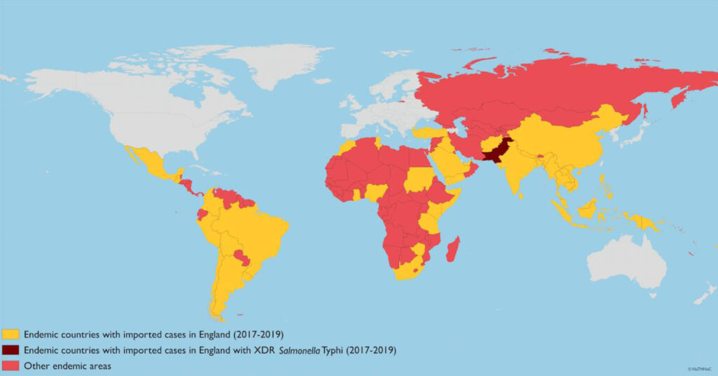
Always treat, even if well and only from stool (cf other salmonella types).
IV ceftriaxone if sepsis/shock, GI bleeding, intestinal perforation, encephalopathy, metastatic infection, total 10-14 days. Else azithromycin (loading dose 1g, 500mg daily 7 days; children 20mg/kg). [UK-PAS] For areas of high resistance and sepsis, Meropenem plus azithro [CDC Yellow Book].
Normal for fever to take 3-5 days to settle. After that, look for persistent locus and consider additional antibiotic. Fever can persist for 10 days with cefalosporins (low intracellular penetrance).
Drug resistance is a big problem. Fluoroquinolone resistance in most of Asia and sub-Saharan Africa. Oral third generation cephalosporins are also effective although inferior in RCTs.
Where nalidixic acid resistant, in vitro susceptibility to quinolones definitely reduced – use of maximum permitted doses and extending treatment course to 10-14 days will result in cure in >90% of cases — although the clinical response is slower.
Lots of data on quinolone use in children, but theoretical risks so Azithromycin preferred although response possibly slower.
=Paediatric autoimmune neuropsychiatric disorders associated with streptococcal infections (PANDAS) – tics and obsessive-compulsive disorder that have an acute onset and relapsing course, related to streptococcal infection.
Swedo first reported 4 cases of acute onset/exacerbation (2 with evidence of strep), treated successfully with immunosuppression, in 1995. Term first suggested in 1998 by National Institute of Mental Health. Presumed to be causative of course, mediated by inflammation. Has ICD code (8E4A) and everything, but still in 2019 “substantial controversy regarding a role for inflammation in tics and OCD”.
Because many children with similar presentations did not have evidence of streptococcus, a broader category of PANS, paediatric acute onset neuropsychiatric syndrome, was created, which is agnostic to cause. But such a broad category, almost unhelpful.
Finding evidence of previous streptococcal infection is not hard, nor uncommon. Tics and OCD are relatively common too. Although evidence of basal ganglia antibodies, these are non-specific. Lots of neurological problems have acute onset, not least epilepsy. 2020 US study found 91.4% of PANDAS cases had at least 1 of 4 neuronal antibodies in the “Cunningham panel”, and most at least 2, cf 32% of normal controls. Levels dropped over time, as did symptoms. CaMKII (synaptic protein) activity higher too. The antibodies studied were:
In a study of 58 children tested with the Cunningham panel, antibody levels dropped over time, and corresponded well with improvement in symptoms (median 1 yr follow up). Nearly all had treatment, 40% had immune therapy (including IVIG, plasmapheresis and rituximab, but not steroids, curiously), antibiotics (about half) or psychotropics. Mental health symptoms did better than movement disorders. A group of patients who had negative initial tests were then positive on repeat testing. https://doi.org/10.1016/j.jneuroim.2019.577138
Neuropsychiatric problems are well recognised in and after Sydenhams chorea so it is not without basis (but other organ systems involved, and good evidence for inflammation). Encephalitis lethargica is an encephalitic illness with parkinsonism, dyskinesias, and psychiatric disturbance as dominant features. It is assumed to be autoimmune. NMDA-R encephalitis is an encephalitis with dramatic psychiatric disturbance, dyskinesias, cognitive alteration, and seizures, associated with autoantibodies against NMDA-R (originally described in relation to ovarian cancer).
Movement disorders are also described associated with systemic lupus erythematosus and antiphospholipid syndrome.
Diagnosis
PANS –
I. Abrupt, dramatic onset of obsessive-compulsive disorder or severely restricted food intake
II. Concurrent presence of additional neuropsychiatric symptoms, (with similarly severe and acute onset), from at least two of the following seven categories:
5. Deterioration in school performance (related to attention-deficit/hyperactivity disorder [ADHD]-like symptoms, memory deficits, cognitive changes)
6. Sensory or motor abnormalities
7. Somatic signs and symptoms, including sleep disturbances, enuresis, or urinary frequency
III. Symptoms are not better explained by a known neurologic or medical disorder, such as Sydenham’s chorea.
So typically very ill, and usually referred urgently to psychiatric services. NB part III – PANS is a “diagnosis of exclusion”. [Swedo, 2012]
Family history is interesting – mental health issues including tic disorders often run in families, but so do autoimmune conditions, and Sydenham’s chorea.
Kiki Chang’s clinical evaluation guideline defines chorea-like movements seen in PANS as “piano playing movements of fingers when arms extended and eyes closed”, which is very specific. Also states this is only valid over age 8?! But then distinguishes this from “full chorea” (not specified), which should prompt search for a different diagnosis.
For PANDAS, specifies history of scarlatiniform rash, impetigo but also perianal/perivulval dermatitis. ASOT should show rise of at least 58% over 4-8 weeks, or else a single reading more than double the upper limit of normal.
Role of Cunningham panel (discussed above) stated as “useful ancillary information” for PANDAS, but unclear role in PANS.
PANDAS Network recommends the following tests, besides usual inflammatory markers:
Vit D, B 12
ANA
Mycoplasma IgA/M
Lyme, EBV, Coxsackie, HHV-6 titres
Treatment
Lack of controlled treatment trial with more than 50 patients.
2 studies looking at exacerbations and throat cultures in Acute onset and non acute onset, non relapsing tics/OCD. V similar patterns of exacerbations and no obvious link to strep infection.
Controlled trials of antibiotics in PANDAS have not provided strong evidence. Large symptom improvements reported in placebo arm too, for example. No evidence for tonsillectomy. 2016 controlled trial of IVIG showed no benefit (n=35, doi: 10.1016/j.jaac.2016.06.017).
Groups often self selected so at risk of bias. No evidence for 4/52 azithromycin in PANS.
2017 PANS/PANDAS guidelines (PANS/PANDAS Consortium) arenot based on high impact journal research, mostly expert opinion. Table 1 lists “treatment approaches” but in the foot notes it states “this is not a definitive treatment algorithm; rather, it is a framework to aid in clinical decision making. Before initiating any of the therapies, clinicians must consider the risk/benefit ratio for their individual patients and provide careful/informed counseling about risk of side effects”.
Parental anxiety can be increased by the theory that “the brain is under inflammatory attack” which also fuels demands for treatment.
Is PANS/PANDAS diagnosis more acceptable than idiopathic psychiatric problem? Perverse incentives to diagnose, investigate and treat, of course. Vaccines get blamed…
Useful perhaps that more research into inflammation getting done, even if putative triggers still unclear. Still v rare however to see high profile research in psychiatric conditions related to inflammation.
Just as interesting to research effect of parental anxiety and expectations on pattern of exacerbations and prognosis.
In response to increasing demands for PANDAS services, the British Paediatric Neurology Association published a consensus statement (2021) that was then criticised by PANDAS groups. The statement does not recommend the use of antibiotics, steroids or IVIG “as they have an insufficient evidence base to say the potential benefits outweigh the risks”, and “their usage could divert the focus away from effective symptom-directed treatments”. When the BPNA then conducted a priority setting exercise, PANDAS came in at number 8. There is now a BPNA PANDAS working group. The latest statement highlights that –
all children presenting with acute onset neuropsychiatric symptoms should receive a full medical evaluation.
referrals from primary care should not be rejected on the basis that a MDT service may not currently be in place, nor on the basis that there are limited RCTs applicable to PANS or PANDAS.
[use] proficiency and judgment, acquired through clinical experience and clinical practice .
note existing international peer-reviewed and published treatment guidelines; discuss with regional and tertiary services.
The most common adverse events of vaccines are fever, local pain or irritation, and local redness or swelling, which are not signs of allergy
With live vaccines, adverse effects can be delayed until 7 to 21 days after immunization; this includes vaccine-induced delayed-onset urticaria, which is commonly mistaken for allergy.
Assessment and allergy-focused clinical history
Testing appropriate for all, whether anaphylaxis, mild symptoms, unknown history or family history.
Gold standard is an oral challenge with a therapeutic dose of amoxicillin, 1hr observation, then 5 day course at home to ensure no delayed reaction.
Low risk individuals, where IgE mediated reaction unlikely, can go direct to challenge without testing.
Puncture and intradermal skin testing is recommended where reaction was within last 12 months, or there were respiratory symptoms.
Skin testing using only penicilloyl-polylysine, with at least 5 mm of wheal and flare greater than wheal as the criteria for a positive test result, is now sufficient to rule out a high risk of having anaphylaxis during a confirmatory oral amoxicillin challenge
Egg is used in the manufacture of a number of vaccines. Whether this is clinically significant or not depends on the vaccine and the severity of the allergy:
MMR – only contraindication is anaphylaxis to MMR or other constituent of MMR vaccine
Influenza – (live nasal or inactivated injectable) patients with “severe anaphylaxis” (ie requiring intensive care) should be vaccinated in hospital.
Varicella, Rabies – no contraindication for egg allergy
Yellow fever – discuss with specialist if egg allergy
Gelatine (derived from pork or beef) can be a cause of allergic reactions esp MMR, Varicella, Japanese encephalitis. Allergy would usually be established from history of food reactions eg gummy/jelly sweets, marshmallows.
Latex – can be present in syringe or bung. See Latex allergy.
Despite these potential causes of allergy, immunisation can often be achieved through graded administration.
Some reports of persistent itchy nodules at injection site due to aluminium delayed hypersensitivity.